当前位置:
X-MOL 学术
›
Eur. J. Neurol.
›
论文详情
Our official English website, www.x-mol.net, welcomes your
feedback! (Note: you will need to create a separate account there.)
Quantitative magnetic resonance imaging of the brachial plexus shows specific changes in nerve architecture in chronic inflammatory demyelinating polyneuropathy, multifocal motor neuropathy and motor neuron disease
European Journal of Neurology ( IF 4.5 ) Pub Date : 2021-05-01 , DOI: 10.1111/ene.14896 Marieke H J van Rosmalen 1, 2 , H Stephan Goedee 1 , Rosina Derks 2 , Fay-Lynn Asselman 1 , Camiel Verhamme 3 , Alberto de Luca 2 , J Hendrikse 2 , W Ludo van der Pol 1 , Martijn Froeling 2
European Journal of Neurology ( IF 4.5 ) Pub Date : 2021-05-01 , DOI: 10.1111/ene.14896 Marieke H J van Rosmalen 1, 2 , H Stephan Goedee 1 , Rosina Derks 2 , Fay-Lynn Asselman 1 , Camiel Verhamme 3 , Alberto de Luca 2 , J Hendrikse 2 , W Ludo van der Pol 1 , Martijn Froeling 2
Affiliation

|
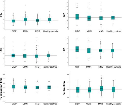
The immunological pathophysiologies of chronic inflammatory demyelinating polyneuropathy (CIDP) and multifocal motor neuropathy (MMN) differ considerably, but neither has been elucidated completely. Quantitative magnetic resonance imaging (MRI) techniques such as diffusion tensor imaging, T2 mapping, and fat fraction analysis may indicate in vivo pathophysiological changes in nerve architecture. Our study aimed to systematically study nerve architecture of the brachial plexus in patients with CIDP, MMN, motor neuron disease (MND) and healthy controls using these quantitative MRI techniques.
中文翻译:

臂丛神经定量磁共振成像显示慢性炎症性脱髓鞘性多发性神经病、多灶性运动神经病和运动神经元疾病中神经结构的特定变化
慢性炎症性脱髓鞘性多发性神经病(CIDP)和多灶性运动神经病(MMN)的免疫病理生理学差异很大,但两者都尚未完全阐明。定量磁共振成像 (MRI) 技术,例如扩散张量成像、T2 映射和脂肪分数分析,可以指示体内神经结构的病理生理变化。我们的研究旨在使用这些定量 MRI 技术系统地研究 CIDP、MMN、运动神经元疾病 (MND) 患者和健康对照患者的臂丛神经结构。
更新日期:2021-05-01
中文翻译:

臂丛神经定量磁共振成像显示慢性炎症性脱髓鞘性多发性神经病、多灶性运动神经病和运动神经元疾病中神经结构的特定变化
慢性炎症性脱髓鞘性多发性神经病(CIDP)和多灶性运动神经病(MMN)的免疫病理生理学差异很大,但两者都尚未完全阐明。定量磁共振成像 (MRI) 技术,例如扩散张量成像、T2 映射和脂肪分数分析,可以指示体内神经结构的病理生理变化。我们的研究旨在使用这些定量 MRI 技术系统地研究 CIDP、MMN、运动神经元疾病 (MND) 患者和健康对照患者的臂丛神经结构。


 京公网安备 11010802027423号