Current Biology ( IF 8.1 ) Pub Date : 2021-04-01 , DOI: 10.1016/j.cub.2021.03.022 Jérémy Magescas 1 , Sani Eskinazi 1 , Michael V Tran 1 , Jessica L Feldman 1

|
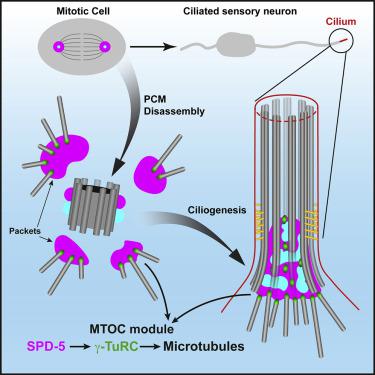
During mitosis in animal cells, the centrosome acts as a microtubule organizing center (MTOC) to assemble the mitotic spindle. MTOC function at the centrosome is driven by proteins within the pericentriolar material (PCM), however the molecular complexity of the PCM makes it difficult to differentiate the proteins required for MTOC activity from other centrosomal functions. We used the natural spatial separation of PCM proteins during mitotic exit to identify a minimal module of proteins required for centrosomal MTOC function in C. elegans. Using tissue-specific degradation, we show that SPD-5, the functional homolog of CDK5RAP2, is essential for embryonic mitosis, while SPD-2/CEP192 and PCMD-1, which are essential in the one-cell embryo, are dispensable. Surprisingly, although the centriole is known to be degraded in the ciliated sensory neurons in C. elegans,1, 2, 3 we find evidence for “centriole-less PCM” at the base of cilia and use this structure as a minimal testbed to dissect centrosomal MTOC function. Super-resolution imaging revealed that this PCM inserts inside the lumen of the ciliary axoneme and directly nucleates the assembly of dendritic microtubules toward the cell body. Tissue-specific degradation in ciliated sensory neurons revealed a role for SPD-5 and the conserved microtubule nucleator γ-TuRC, but not SPD-2 or PCMD-1, in MTOC function at centriole-less PCM. This MTOC function was in the absence of regulation by mitotic kinases, highlighting the intrinsic ability of these proteins to drive microtubule growth and organization and further supporting a model that SPD-5 is the primary driver of MTOC function at the PCM.
中文翻译:

无中心粒的中心粒周围材料作为秀丽隐杆线虫感觉纤毛底部的微管组织中心
在动物细胞的有丝分裂过程中,中心体充当微管组织中心 (MTOC) 来组装有丝分裂纺锤体。中心体的 MTOC 功能由中心体周围材料 (PCM) 中的蛋白质驱动,但是 PCM 的分子复杂性使得难以区分 MTOC 活性所需的蛋白质与其他中心体功能。我们在有丝分裂退出期间使用 PCM 蛋白的自然空间分离来确定秀丽隐杆线虫中心体 MTOC 功能所需的最小蛋白质模块。使用组织特异性降解,我们表明 CDK5RAP2 的功能同源物 SPD-5 对胚胎有丝分裂至关重要,而在单细胞胚胎中必不可少的 SPD-2/CEP192 和 PCMD-1 是可有可无的。令人惊讶的是,尽管已知中心粒在秀丽隐杆线虫的纤毛感觉神经元中被降解,1,2,3 我们在纤毛底部发现了“无中心粒 PCM”的证据,并将该结构用作解剖中心体 MTOC 功能的最小试验台。超分辨率成像显示,这种 PCM 插入睫状轴丝的管腔内,并直接使树突状微管的组装成核朝向细胞体。纤毛感觉神经元的组织特异性降解揭示了 SPD-5 和保守的微管成核因子 γ-TuRC,但不是 SPD-2 或 PCMD-1,在无中心粒 PCM 的 MTOC 功能中的作用。这种 MTOC 功能缺乏有丝分裂激酶的调节,突出了这些蛋白质驱动微管生长和组织的内在能力,并进一步支持了 SPD-5 是 PCM 中 MTOC 功能的主要驱动因素的模型。


 京公网安备 11010802027423号