Our official English website, www.x-mol.net, welcomes your
feedback! (Note: you will need to create a separate account there.)
Multiscale structural characterization of the vertebral endplate in animal models
Journal of Anatomy ( IF 1.8 ) Pub Date : 2021-01-31 , DOI: 10.1111/joa.13402
Magdalena Wojtków 1 , Maciej Głowacki 2 , Celina Pezowicz 1
Journal of Anatomy ( IF 1.8 ) Pub Date : 2021-01-31 , DOI: 10.1111/joa.13402
Magdalena Wojtków 1 , Maciej Głowacki 2 , Celina Pezowicz 1
Affiliation
|
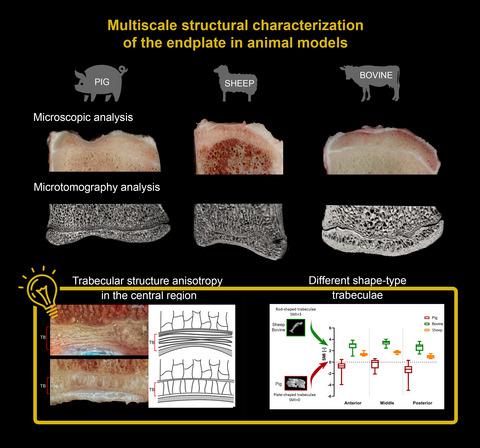
Research in the field of spinal biomechanics, including analyses of the impact of implants on the stability of the spine, is conducted extensively in animal models. One of the basic problems in spinal implantation is the transfer and distribution of loads carried by the spine on the surfaces of the vertebral bodies. An important factor in proper cooperation of spinal implants with the vertebrae is the endplate (EP), which is why the EP in the animal model used for testing should be as similar as possible to the human EP. Therefore, this study involved multiscale structural and morphometric analyses of the animal models most commonly used in spinal biomechanics research, i.e. pig, ovine, and bovine tail. The tests were performed on 28 lumbar porcine, ovine, and bovine vertebrae. Both cranial and caudal EPs were analysed in three selected areas: anterior, middle, and posterior EPs. The conducted tests included a morphometric analysis of the trabecular bone (TB) layer of the EP as well as microscopic analysis at the mesoscale (total thickness) and microscale (thickness of the individual EP layers). The porcine EP had a characteristic increased circumferential thickness (~3 mm) with a significant narrowing in the central region (50%–60%). The convex cranial ovine EP had a constant thickness throughout the cross-section and the concave caudal EP showed ~35% narrowing in the central region. The thickest EPs were observed in the bovine tail model with negligibly small narrowing in the central region (~5%). The thickness of the cartilaginous layer in the porcine and bovine models reached up to 1 mm in the peripheral regions and decreased in the central part. The growth plate layer had a similar thickness in all the models. On the other hand, the narrowing of the total thickness of the EPs in the central region was mainly due to a decrease in the VEP thickness. In the ovine and bovine models, the central region of the EP was characterized by large isotropy and trabeculae of mixed or rod-like shape. By contrast, in the pig, this region had plate-like trabeculae of anisotropic nature. The porcine model was identified as best reflecting the shape and structure of the human EP and as the best surrogate model for the human EP model. This choice is particularly important in the context of biomechanical research.
中文翻译:

动物模型中椎体终板的多尺度结构表征
脊柱生物力学领域的研究,包括分析植入物对脊柱稳定性的影响,在动物模型中广泛进行。脊柱植入的基本问题之一是脊柱所承载的负载在椎体表面上的转移和分布。脊柱植入物与椎骨正确配合的一个重要因素是终板 (EP),这就是为什么用于测试的动物模型中的 EP 应尽可能与人类 EP 相似。因此,本研究涉及对脊柱生物力学研究中最常用的动物模型(即猪、绵羊和牛尾)进行多尺度结构和形态测量分析。这些测试是在 28 块猪、羊和牛的腰椎上进行的。在三个选定区域对颅侧和尾侧 EP 进行了分析:前、中、后 EP。进行的测试包括 EP 骨小梁 (TB) 层的形态测量分析以及中尺度(总厚度)和微观尺度(各个 EP 层的厚度)的显微分析。猪 EP 的特征是周向厚度增加(约 3 毫米),而中心区域显着变窄(50%–60%)。凸面绵羊 EP 在整个横截面中具有恒定的厚度,而凹面尾部 EP 在中心区域显示约 35% 的狭窄。在牛尾模型中观察到最厚的 EP,中央区域有可忽略不计的小狭窄 (~5%)。猪和牛模型的软骨层厚度在周边区域达到1毫米,而在中心部分减少。所有模型中的生长板层具有相似的厚度。另一方面,中心区域的EP总厚度变窄主要是由于VEP厚度的减小。在绵羊和牛模型中,EP 中心区域的特征是大的各向同性和混合或杆状形状的小梁。相比之下,在猪中,该区域具有各向异性的板状小梁。猪模型被认为最能反映人类EP的形状和结构,并且是人类EP模型的最佳替代模型。这种选择在生物力学研究的背景下尤其重要。在绵羊和牛模型中,EP 中心区域的特征是大的各向同性和混合或杆状形状的小梁。相比之下,在猪中,该区域具有各向异性的板状小梁。猪模型被认为最能反映人类EP的形状和结构,并且是人类EP模型的最佳替代模型。这种选择在生物力学研究的背景下尤其重要。在绵羊和牛模型中,EP 中心区域的特征是大的各向同性和混合或杆状形状的小梁。相比之下,在猪中,该区域具有各向异性的板状小梁。猪模型被认为最能反映人类EP的形状和结构,并且是人类EP模型的最佳替代模型。这种选择在生物力学研究的背景下尤其重要。
更新日期:2021-01-31
中文翻译:
动物模型中椎体终板的多尺度结构表征
脊柱生物力学领域的研究,包括分析植入物对脊柱稳定性的影响,在动物模型中广泛进行。脊柱植入的基本问题之一是脊柱所承载的负载在椎体表面上的转移和分布。脊柱植入物与椎骨正确配合的一个重要因素是终板 (EP),这就是为什么用于测试的动物模型中的 EP 应尽可能与人类 EP 相似。因此,本研究涉及对脊柱生物力学研究中最常用的动物模型(即猪、绵羊和牛尾)进行多尺度结构和形态测量分析。这些测试是在 28 块猪、羊和牛的腰椎上进行的。在三个选定区域对颅侧和尾侧 EP 进行了分析:前、中、后 EP。进行的测试包括 EP 骨小梁 (TB) 层的形态测量分析以及中尺度(总厚度)和微观尺度(各个 EP 层的厚度)的显微分析。猪 EP 的特征是周向厚度增加(约 3 毫米),而中心区域显着变窄(50%–60%)。凸面绵羊 EP 在整个横截面中具有恒定的厚度,而凹面尾部 EP 在中心区域显示约 35% 的狭窄。在牛尾模型中观察到最厚的 EP,中央区域有可忽略不计的小狭窄 (~5%)。猪和牛模型的软骨层厚度在周边区域达到1毫米,而在中心部分减少。所有模型中的生长板层具有相似的厚度。另一方面,中心区域的EP总厚度变窄主要是由于VEP厚度的减小。在绵羊和牛模型中,EP 中心区域的特征是大的各向同性和混合或杆状形状的小梁。相比之下,在猪中,该区域具有各向异性的板状小梁。猪模型被认为最能反映人类EP的形状和结构,并且是人类EP模型的最佳替代模型。这种选择在生物力学研究的背景下尤其重要。在绵羊和牛模型中,EP 中心区域的特征是大的各向同性和混合或杆状形状的小梁。相比之下,在猪中,该区域具有各向异性的板状小梁。猪模型被认为最能反映人类EP的形状和结构,并且是人类EP模型的最佳替代模型。这种选择在生物力学研究的背景下尤其重要。在绵羊和牛模型中,EP 中心区域的特征是大的各向同性和混合或杆状形状的小梁。相比之下,在猪中,该区域具有各向异性的板状小梁。猪模型被认为最能反映人类EP的形状和结构,并且是人类EP模型的最佳替代模型。这种选择在生物力学研究的背景下尤其重要。


 京公网安备 11010802027423号