当前位置:
X-MOL 学术
›
Atherosclerosis
›
论文详情
Our official English website, www.x-mol.net, welcomes your
feedback! (Note: you will need to create a separate account there.)
Vascular imaging of atherosclerosis: Strengths and weaknesses
Atherosclerosis ( IF 4.9 ) Pub Date : 2021-02-01 , DOI: 10.1016/j.atherosclerosis.2020.12.021 Laura E Mantella 1 , Kiera Liblik 2 , Amer M Johri 3
Atherosclerosis ( IF 4.9 ) Pub Date : 2021-02-01 , DOI: 10.1016/j.atherosclerosis.2020.12.021 Laura E Mantella 1 , Kiera Liblik 2 , Amer M Johri 3
Affiliation

|
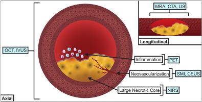
Atherosclerosis is an inflammatory disease that can lead to several complications such as ischemic heart disease, stroke, and peripheral vascular disease. Therefore, researchers and clinicians rely heavily on the use of imaging modalities to identify, and more recently, quantify the burden of atherosclerosis in the aorta, carotid arteries, coronary arteries, and peripheral vasculature. These imaging techniques vary in invasiveness, cost, resolution, radiation exposure, and presence of artifacts. Consequently, a detailed understanding of the risks and benefits of each technique is crucial prior to their introduction into routine cardiovascular screening. Additionally, recent research in the field of microvascular imaging has proven to be important in the field of atherosclerosis. Using techniques such as contrast-enhanced ultrasound and superb microvascular imaging, researchers have been able to detect blood vessels within a plaque lesion that may contribute to vulnerability and rupture. This paper will review the strengths and weaknesses of the various imaging techniques used to measure atherosclerotic burden. Furthermore, it will discuss the future of advanced imaging modalities as potential biomarkers for atherosclerosis.
中文翻译:

动脉粥样硬化的血管成像:优点和缺点
动脉粥样硬化是一种炎症性疾病,可导致多种并发症,如缺血性心脏病、中风和外周血管疾病。因此,研究人员和临床医生在很大程度上依赖于使用成像方式来识别和量化主动脉、颈动脉、冠状动脉和外周脉管系统中动脉粥样硬化的负担。这些成像技术在侵入性、成本、分辨率、辐射暴露和伪影的存在方面各不相同。因此,在将每种技术引入常规心血管筛查之前,详细了解每种技术的风险和益处至关重要。此外,最近在微血管成像领域的研究已证明在动脉粥样硬化领域很重要。使用对比增强超声和精湛的微血管成像等技术,研究人员已经能够检测出斑块病变内可能导致脆弱和破裂的血管。本文将回顾用于测量动脉粥样硬化负担的各种成像技术的优缺点。此外,它将讨论作为动脉粥样硬化潜在生物标志物的先进成像方式的未来。
更新日期:2021-02-01
中文翻译:

动脉粥样硬化的血管成像:优点和缺点
动脉粥样硬化是一种炎症性疾病,可导致多种并发症,如缺血性心脏病、中风和外周血管疾病。因此,研究人员和临床医生在很大程度上依赖于使用成像方式来识别和量化主动脉、颈动脉、冠状动脉和外周脉管系统中动脉粥样硬化的负担。这些成像技术在侵入性、成本、分辨率、辐射暴露和伪影的存在方面各不相同。因此,在将每种技术引入常规心血管筛查之前,详细了解每种技术的风险和益处至关重要。此外,最近在微血管成像领域的研究已证明在动脉粥样硬化领域很重要。使用对比增强超声和精湛的微血管成像等技术,研究人员已经能够检测出斑块病变内可能导致脆弱和破裂的血管。本文将回顾用于测量动脉粥样硬化负担的各种成像技术的优缺点。此外,它将讨论作为动脉粥样硬化潜在生物标志物的先进成像方式的未来。


 京公网安备 11010802027423号