Neuron ( IF 14.7 ) Pub Date : 2020-12-07 , DOI: 10.1016/j.neuron.2020.11.011 Jennifer D. Whitesell , Adam Liska , Ludovico Coletta , Karla E. Hirokawa , Phillip Bohn , Ali Williford , Peter A. Groblewski , Nile Graddis , Leonard Kuan , Joseph E. Knox , Anh Ho , Wayne Wakeman , Philip R. Nicovich , Thuc Nghi Nguyen , Cindy T.J. van Velthoven , Emma Garren , Olivia Fong , Maitham Naeemi , Alex M. Henry , Nick Dee , Kimberly A. Smith , Boaz Levi , David Feng , Lydia Ng , Bosiljka Tasic , Hongkui Zeng , Stefan Mihalas , Alessandro Gozzi , Julie A. Harris

|
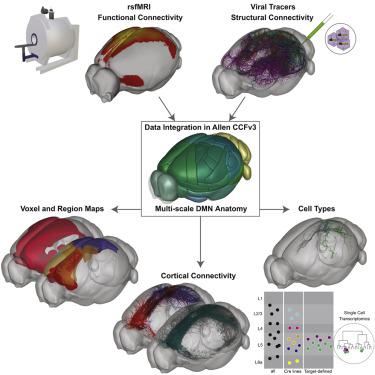
The evolutionarily conserved default mode network (DMN) is a distributed set of brain regions coactivated during resting states that is vulnerable to brain disorders. How disease affects the DMN is unknown, but detailed anatomical descriptions could provide clues. Mice offer an opportunity to investigate structural connectivity of the DMN across spatial scales with cell-type resolution. We co-registered maps from functional magnetic resonance imaging and axonal tracing experiments into the 3D Allen mouse brain reference atlas. We find that the mouse DMN consists of preferentially interconnected cortical regions. As a population, DMN layer 2/3 (L2/3) neurons project almost exclusively to other DMN regions, whereas L5 neurons project in and out of the DMN. In the retrosplenial cortex, a core DMN region, we identify two L5 projection types differentiated by in- or out-DMN targets, laminar position, and gene expression. These results provide a multi-scale description of the anatomical correlates of the mouse DMN.
中文翻译:

鼠标默认模式网络的区域,层和特定于单元格类型的连接
进化保守的默认模式网络(DMN)是一组分布的大脑区域,在休息状态期间容易受到激活,容易受到脑部疾病的影响。疾病如何影响DMN尚不清楚,但是详细的解剖描述可能会提供线索。小鼠提供了一个机会来研究具有细胞类型分辨率的DMN在空间尺度上的结构连通性。我们将功能磁共振成像和轴突跟踪实验中的地图共同注册到3D Allen小鼠大脑参考地图集中。我们发现,鼠标DMN由优先互连的皮质区域组成。作为种群,DMN第2/3(L2 / 3)层神经元几乎只投射到其他DMN区域,而L5神经元则投射到DMN内外。在脾后皮质,DMN核心区域,我们确定了两种L5投影类型,它们分别由入-出-DMN靶,层流位置和基因表达来区分。这些结果提供了鼠标DMN的解剖相关性的多尺度描述。


 京公网安备 11010802027423号