Acta Biomaterialia ( IF 9.4 ) Pub Date : 2020-10-21 , DOI: 10.1016/j.actbio.2020.10.028 Jingnan Zhang , Renping Zhao , Bin Li , Aleeza Farrukh , Markus Hoth , Bin Qu , Aránzazu del Campo

|
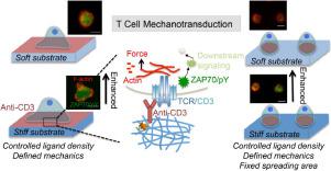
The analysis of T cell responses to mechanical properties of antigen presenting cells (APC) is experimentally challenging at T cell-APC interfaces. Soft hydrogels with adjustable mechanical properties and biofunctionalization are useful reductionist models to address this problem. Here, we report a methodology to fabricate micropatterned soft hydrogels with defined stiffness to form spatially confined T cell/hydrogel contact interfaces at micrometer scale. Using automatized microcontact printing we prepared arrays of anti-CD3 microdots on poly(acrylamide) hydrogels with Young's Modulus in the range of 2 to 50 kPa. We optimized the printing process to obtain anti-CD3 microdots with constant area (50 µm2, corresponding to 8 µm diameter) and comparable anti-CD3 density on hydrogels of different stiffness. The anti-CD3 arrays were recognized by T cells and restricted cell attachment to the printed areas. To test functionality of the hydrogel-T cell contact, we analyzed several key events downstream of T cell receptor (TCR) activation. Anti-CD3 arrays on hydrogels activated calcium influx, induced rearrangement of the actin cytoskeleton, and led to Zeta-chain-associated protein kinase 70 (ZAP70) phosphorylation. Interestingly, upon increase in the stiffness, ZAP70 phosphorylation was enhanced, whereas the rearrangements of F-actin (F-actin clearance) and phosphorylated ZAP70 (ZAP70/pY centralization) were unaffected. Our results show that micropatterned hydrogels allow tuning of stiffness and receptor presentation to analyze TCR mediated T cell activation as function of mechanical, biochemical, and geometrical parameters.
中文翻译:

微模式的软水凝胶可研究受体和作用于T细胞活化的相互作用
T细胞对抗原呈递细胞(APC)的机械性能的响应分析在T细胞-APC界面上具有挑战性。具有可调节机械性能和生物功能化的软水凝胶是解决此问题的有用的还原剂模型。在这里,我们报告了一种方法,可以制造具有确定的刚度的微图案软水凝胶,以形成微米级的空间受限的T细胞/水凝胶接触界面。使用自动微接触印刷,我们在杨氏模量范围为2至50 kPa的聚丙烯酰胺水凝胶上制备了抗CD3微粒阵列。我们优化了打印过程,以获得具有恒定面积(50 µm 2)的抗CD3微点,对应于8 µm直径),并在不同硬度的水凝胶上具有可比的抗CD3密度。抗CD3阵列可被T细胞识别并限制细胞附着到印刷区域。为了测试水凝胶-T细胞接触的功能,我们分析了T细胞受体(TCR)激活下游的几个关键事件。水凝胶上的抗CD3阵列激活钙内流,诱导肌动蛋白细胞骨架重排,并导致与Zeta链相关的蛋白激酶70(ZAP70)磷酸化。有趣的是,刚度增加时,ZAP70的磷酸化增强,而F-肌动蛋白(F-肌动蛋白清除率)和磷酸化ZAP70(ZAP70 / pY中心化)的重排不受影响。


 京公网安备 11010802027423号