Our official English website, www.x-mol.net, welcomes your
feedback! (Note: you will need to create a separate account there.)
Contrast-enhanced micro-computed tomography of articular cartilage morphology with ioversol and iomeprol.
Journal of Anatomy ( IF 1.8 ) Pub Date : 2020-07-19 , DOI: 10.1111/joa.13271 Colet E M Ter Voert 1 , R Y Nigel Kour 1 , Bente C J van Teeffelen 1 , Niloufar Ansari 1 , Kathryn S Stok 1
Journal of Anatomy ( IF 1.8 ) Pub Date : 2020-07-19 , DOI: 10.1111/joa.13271 Colet E M Ter Voert 1 , R Y Nigel Kour 1 , Bente C J van Teeffelen 1 , Niloufar Ansari 1 , Kathryn S Stok 1
Affiliation

|
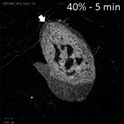
Non‐ionic, low‐osmolar contrast agents (CAs) used for computed tomography, such as Optiray (ioversol) and Iomeron (iomeprol), are associated with the reduced risk of adverse reactions and toxicity in comparison with ionic CAs, such as Hexabrix. Hexabrix has previously been used for imaging articular cartilage but has been commercially discontinued. This study aimed to evaluate the efficacy of Optiray and Iomeron as alternatives for visualisation of articular cartilage in small animal joints using contrast‐enhanced micro‐computed tomography (CECT). For this purpose, mouse femora were immersed in different concentrations (20%–50%) of Optiray 350 or Iomeron 350 for periods of time starting at five minutes. The femoral condyles were scanned ex vivo using CECT, and regions of articular cartilage manually contoured to calculate mean attenuation at each time point and concentration. For both CAs, a 30% CA concentration produced a mean cartilage attenuation optimally distinct from both bone and background signal, whilst 5‐min immersion times were sufficient for equilibration of CA absorption. Additionally, plugs of bovine articular cartilage were digested by chondroitinase ABC to produce a spectrum of glycosaminoglycan (GAG) content. These samples were immersed in CA and assessed for any correlation between mean attenuation and GAG content. No significant correlation was found between attenuation and cartilage GAG content for either CAs. In conclusion, Optiray and Iomeron enable high‐resolution morphological assessment of articular cartilage in small animals using CECT; however, they are not indicative of GAG content.
中文翻译:

使用碘佛醇和碘美普尔对关节软骨形态进行对比增强显微计算机断层扫描。
与 Hexabrix 等离子型 CA 相比,用于计算机断层扫描的非离子型低渗造影剂 (CA),例如 Optiray (ioversol) 和 Iomeron (iomeprol),与不良反应和毒性风险降低有关。Hexabrix 以前曾用于关节软骨成像,但已停产。本研究旨在评估 Optiray 和 Iomeron 作为使用对比增强显微计算机断层扫描 (CECT) 对小动物关节中关节软骨进行可视化的替代方案的功效。为此,将小鼠股骨浸入不同浓度 (20%–50%) 的 Optiray 350 或 Iomeron 350 中,从五分钟开始持续一段时间。使用 CECT 离体扫描股骨髁,和关节软骨区域手动绘制轮廓以计算每个时间点和浓度的平均衰减。对于这两种 CA,30% 的 CA 浓度产生了与骨骼和背景信号最佳不同的平均软骨衰减,而 5 分钟的浸泡时间足以平衡 CA 吸收。此外,牛关节软骨栓被软骨素酶 ABC 消化,产生一系列糖胺聚糖 (GAG) 含量。将这些样品浸入 CA 中并评估平均衰减和 GAG 含量之间的任何相关性。对于任一 CA,在衰减和软骨 GAG 含量之间未发现显着相关性。总之,Optiray 和 Iomeron 能够使用 CECT 对小动物的关节软骨进行高分辨率形态学评估;然而,
更新日期:2020-07-19
中文翻译:

使用碘佛醇和碘美普尔对关节软骨形态进行对比增强显微计算机断层扫描。
与 Hexabrix 等离子型 CA 相比,用于计算机断层扫描的非离子型低渗造影剂 (CA),例如 Optiray (ioversol) 和 Iomeron (iomeprol),与不良反应和毒性风险降低有关。Hexabrix 以前曾用于关节软骨成像,但已停产。本研究旨在评估 Optiray 和 Iomeron 作为使用对比增强显微计算机断层扫描 (CECT) 对小动物关节中关节软骨进行可视化的替代方案的功效。为此,将小鼠股骨浸入不同浓度 (20%–50%) 的 Optiray 350 或 Iomeron 350 中,从五分钟开始持续一段时间。使用 CECT 离体扫描股骨髁,和关节软骨区域手动绘制轮廓以计算每个时间点和浓度的平均衰减。对于这两种 CA,30% 的 CA 浓度产生了与骨骼和背景信号最佳不同的平均软骨衰减,而 5 分钟的浸泡时间足以平衡 CA 吸收。此外,牛关节软骨栓被软骨素酶 ABC 消化,产生一系列糖胺聚糖 (GAG) 含量。将这些样品浸入 CA 中并评估平均衰减和 GAG 含量之间的任何相关性。对于任一 CA,在衰减和软骨 GAG 含量之间未发现显着相关性。总之,Optiray 和 Iomeron 能够使用 CECT 对小动物的关节软骨进行高分辨率形态学评估;然而,


 京公网安备 11010802027423号