Our official English website, www.x-mol.net, welcomes your
feedback! (Note: you will need to create a separate account there.)
Three‐dimensional reconstruction of the luminal structure of human seminal vesicle
Journal of Anatomy ( IF 1.8 ) Pub Date : 2020-07-13 , DOI: 10.1111/joa.13269 Je-Sung Lee 1 , In-Seung Yeo 1 , Hye-In Lee 1 , Jung-Ah Park 1 , Ki-Seok Koh 1 , Wu-Chul Song 1
Journal of Anatomy ( IF 1.8 ) Pub Date : 2020-07-13 , DOI: 10.1111/joa.13269 Je-Sung Lee 1 , In-Seung Yeo 1 , Hye-In Lee 1 , Jung-Ah Park 1 , Ki-Seok Koh 1 , Wu-Chul Song 1
Affiliation

|
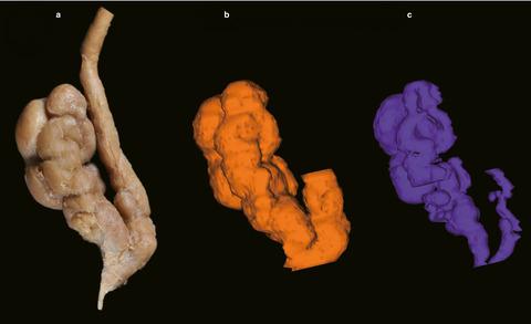
The seminal vesicles are the glands of male reproductive organs that produce the fluid and nutrient constituents of semen. It has been believed for a long time that the lumen of a seminal vesicle was a single‐coiled tubular structure with irregular diverticula. There are several previous reports on the symmetry, differences in morphological sizes and classification of the seminal vesicles. However, a three‐dimensional‐coiled tubular structure is difficult to understand using a classical anatomical methodology, and hence, three‐dimensional reconstruction is needed to understand the structure of the lumen. Thirty‐one seminal vesicles harvested from 21 formalin‐embalmed cadavers were investigated. The seminal vesicle along with the ampulla of the ductus deferens was separated, and the length and width of each seminal vesicle were measured. The vesicles were then embedded in coloured paraffin, and the resulting paraffin block was sectioned transversely and photographed at an interval of 500 μm, with the sectioned surfaces then utilized in three‐dimensional reconstruction performed by ‘Reconstruct’ software. The mean length and width of the seminal vesicles were 39.4 mm and 13.4 mm, respectively, and the right seminal vesicle was a little larger than the one on the left. The size differed from previous reports, while the luminal structure was similar to the classification of Aboul‐azm (Archives of Andrology, 3, 1979, 287–292) but differed from that of Pereira (AJR. American Journal of Roentgenology, 69, 1953, 361–379). The seminal vesicles typically comprised about 9 curls and had about 12 diverticula. The seminal vesicles resembled a skein of coral rather than comprising a single strand. These findings will help in improving the understanding of pathophysiologies of the seminal vesicles, such as recurrent inflammation of the gland.
中文翻译:

人精囊管腔结构的三维重建
精囊是男性生殖器官的腺体,可产生精液的液体和营养成分。长期以来,人们一直认为精囊的腔是一个单盘管状结构,带有不规则的憩室。以前有几篇关于精囊的对称性、形态大小差异和分类的报道。然而,三维螺旋管状结构很难用经典的解剖学方法来理解,因此,需要三维重建来理解管腔的结构。对从 21 具福尔马林防腐尸体中采集的 31 个精囊进行了研究。分离输精管壶腹和精囊,测量每个精囊的长度和宽度。然后将囊泡包埋在彩色石蜡中,将所得石蜡块横向切片并以 500 μm 的间隔拍照,然后将切片表面用于“重建”软件执行的三维重建。精囊的平均长度和宽度分别为 39.4 mm 和 13.4 mm,右侧精囊略大于左侧。大小与之前的报告不同,而管腔结构类似于 Aboul-azm 的分类(Archives of Andrology, 3, 1979, 287–292),但与 Pereira 的分类不同(AJR. American Journal of Roentgenology, 69, 1953 , 361–379). 精囊通常包含约 9 个卷曲并具有约 12 个憩室。精囊类似于一束珊瑚,而不是单链。这些发现将有助于提高对精囊病理生理学的理解,例如腺体的复发性炎症。
更新日期:2020-07-13
中文翻译:

人精囊管腔结构的三维重建
精囊是男性生殖器官的腺体,可产生精液的液体和营养成分。长期以来,人们一直认为精囊的腔是一个单盘管状结构,带有不规则的憩室。以前有几篇关于精囊的对称性、形态大小差异和分类的报道。然而,三维螺旋管状结构很难用经典的解剖学方法来理解,因此,需要三维重建来理解管腔的结构。对从 21 具福尔马林防腐尸体中采集的 31 个精囊进行了研究。分离输精管壶腹和精囊,测量每个精囊的长度和宽度。然后将囊泡包埋在彩色石蜡中,将所得石蜡块横向切片并以 500 μm 的间隔拍照,然后将切片表面用于“重建”软件执行的三维重建。精囊的平均长度和宽度分别为 39.4 mm 和 13.4 mm,右侧精囊略大于左侧。大小与之前的报告不同,而管腔结构类似于 Aboul-azm 的分类(Archives of Andrology, 3, 1979, 287–292),但与 Pereira 的分类不同(AJR. American Journal of Roentgenology, 69, 1953 , 361–379). 精囊通常包含约 9 个卷曲并具有约 12 个憩室。精囊类似于一束珊瑚,而不是单链。这些发现将有助于提高对精囊病理生理学的理解,例如腺体的复发性炎症。


 京公网安备 11010802027423号