Our official English website, www.x-mol.net, welcomes your
feedback! (Note: you will need to create a separate account there.)
Anatomy of the female pelvic nerves: a macroscopic study of the hypogastric plexus and their relations and variations.
Journal of Anatomy ( IF 1.8 ) Pub Date : 2020-05-19 , DOI: 10.1111/joa.13206
Valerie Aurore 1 , Raphael Röthlisberger 1, 2 , Nane Boemke 1 , Ruslan Hlushchuk 1 , Hannes Bangerter 1 , Mathias Bergmann 1 , Sara Imboden 3 , Michael D Mueller 3 , Elisabeth Eppler 1 , Valentin Djonov 1
Journal of Anatomy ( IF 1.8 ) Pub Date : 2020-05-19 , DOI: 10.1111/joa.13206
Valerie Aurore 1 , Raphael Röthlisberger 1, 2 , Nane Boemke 1 , Ruslan Hlushchuk 1 , Hannes Bangerter 1 , Mathias Bergmann 1 , Sara Imboden 3 , Michael D Mueller 3 , Elisabeth Eppler 1 , Valentin Djonov 1
Affiliation

|
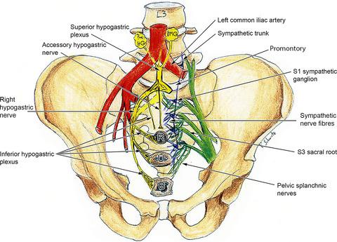
The autonomic nerves of the lesser pelvis are particularly prone to iatrogenic lesions due to their exposed position during manifold surgical interventions. Nevertheless, the cause of rectal and urinary incontinence or sexual dysfunctions, for example after rectal cancer resection or hysterectomy, remains largely understudied, particularly with regard to the female pelvic autonomic plexuses. This study focused on the macroscopic description of the superior hypogastric plexus, hypogastric nerves, inferior hypogastric plexus, the parasympathetic pelvic splanchnic nerves and the sympathetic fibres. Their arrangement is described in relation to commonly used surgical landmarks such as the sacral promontory, ureters, uterosacral ligaments, uterine and rectal blood vessels. Thirty‐one embalmed female pelvises from 20 formalin‐fixed and 11 Thiel‐fixed cadavers were prepared. In all cases explored, the superior hypogastric plexus was situated anterior to the bifurcation of the abdominal aorta. In 60% of specimens, it reached the sacral promontory, whereas in 40% of specimens, it continued across the pelvic brim until S1. In about 25% of the subjects, we detected an accessory hypogastric nerve, which has not been systematically described so far. It originated medially from the inferior margin of the superior hypogastric plexus and continued medially into the presacral space. The existence of an accessory hypogastric nerve was confirmed during laparoscopy and by histological examination. The inferior hypogastric plexuses formed fan‐shaped plexiform structures at the end of both hypogastric nerves, exactly at the junction of the ureter and the posterior wall of the uterine artery at the uterosacral ligament. In addition to the pelvic splanchnic nerves from S2–S4, which joined the inferior hypogastric plexus, 18% of the specimens in the present study revealed an additional pelvic splanchnic nerve originating from the S1 sacral root. In general, form, breadth and alignment of the autonomic nerves displayed large individual variations, which could also have a clinical impact on the postoperative function of the pelvic organs. The study serves as a basis for future investigations on the autonomic innervation of the female pelvic organs.
中文翻译:

女性骨盆神经的解剖:下腹神经丛及其关系和变化的宏观研究。
小骨盆的自主神经由于在多种手术干预过程中的暴露位置而特别容易出现医源性损伤。然而,直肠和尿失禁或性功能障碍的原因,例如在直肠癌切除术或子宫切除术后,仍然很大程度上未被研究,特别是关于女性盆腔自主神经丛。本研究侧重于对上腹下丛、下腹神经、下腹下丛、副交感盆腔内脏神经和交感神经纤维的宏观描述。它们的排列与常用的手术标志有关,如骶岬、输尿管、子宫骶韧带、子宫和直肠血管。从 20 具福尔马林固定和 11 具泰尔固定的尸体中制备了 31 具防腐处理的女性骨盆。在所有探索的病例中,上腹下神经丛都位于腹主动脉分叉的前面。在 60% 的标本中,它到达了骶岬,而在 40% 的标本中,它继续穿过骨盆边缘直到 S1。在大约 25% 的受试者中,我们检测到了一条腹下副神经,目前尚未系统地描述该神经。它从上腹下神经丛的下缘向内侧起源并继续向内侧进入骶前空间。腹腔镜检查和组织学检查证实了腹下副神经的存在。下腹下丛在两条下腹神经末端形成扇形丛状结构,正好在输尿管和子宫骶韧带处子宫动脉后壁的交界处。除了来自 S2-S4 的骨盆内脏神经,它与下腹下丛相连,本研究中 18% 的标本显示额外的骨盆内脏神经起源于 S1 骶根。一般来说,自主神经的形态、宽度和排列表现出较大的个体差异,这也可能对盆腔器官的术后功能产生临床影响。该研究为未来研究女性盆腔器官的自主神经支配奠定了基础。本研究中 18% 的标本显示额外的骨盆内脏神经起源于 S1 骶根。一般来说,自主神经的形态、宽度和排列表现出较大的个体差异,这也可能对盆腔器官的术后功能产生临床影响。该研究为未来研究女性盆腔器官的自主神经支配奠定了基础。本研究中 18% 的标本显示额外的骨盆内脏神经起源于 S1 骶根。一般来说,自主神经的形态、宽度和排列表现出较大的个体差异,这也可能对盆腔器官的术后功能产生临床影响。该研究为未来研究女性盆腔器官的自主神经支配奠定了基础。
更新日期:2020-05-19
中文翻译:

女性骨盆神经的解剖:下腹神经丛及其关系和变化的宏观研究。
小骨盆的自主神经由于在多种手术干预过程中的暴露位置而特别容易出现医源性损伤。然而,直肠和尿失禁或性功能障碍的原因,例如在直肠癌切除术或子宫切除术后,仍然很大程度上未被研究,特别是关于女性盆腔自主神经丛。本研究侧重于对上腹下丛、下腹神经、下腹下丛、副交感盆腔内脏神经和交感神经纤维的宏观描述。它们的排列与常用的手术标志有关,如骶岬、输尿管、子宫骶韧带、子宫和直肠血管。从 20 具福尔马林固定和 11 具泰尔固定的尸体中制备了 31 具防腐处理的女性骨盆。在所有探索的病例中,上腹下神经丛都位于腹主动脉分叉的前面。在 60% 的标本中,它到达了骶岬,而在 40% 的标本中,它继续穿过骨盆边缘直到 S1。在大约 25% 的受试者中,我们检测到了一条腹下副神经,目前尚未系统地描述该神经。它从上腹下神经丛的下缘向内侧起源并继续向内侧进入骶前空间。腹腔镜检查和组织学检查证实了腹下副神经的存在。下腹下丛在两条下腹神经末端形成扇形丛状结构,正好在输尿管和子宫骶韧带处子宫动脉后壁的交界处。除了来自 S2-S4 的骨盆内脏神经,它与下腹下丛相连,本研究中 18% 的标本显示额外的骨盆内脏神经起源于 S1 骶根。一般来说,自主神经的形态、宽度和排列表现出较大的个体差异,这也可能对盆腔器官的术后功能产生临床影响。该研究为未来研究女性盆腔器官的自主神经支配奠定了基础。本研究中 18% 的标本显示额外的骨盆内脏神经起源于 S1 骶根。一般来说,自主神经的形态、宽度和排列表现出较大的个体差异,这也可能对盆腔器官的术后功能产生临床影响。该研究为未来研究女性盆腔器官的自主神经支配奠定了基础。本研究中 18% 的标本显示额外的骨盆内脏神经起源于 S1 骶根。一般来说,自主神经的形态、宽度和排列表现出较大的个体差异,这也可能对盆腔器官的术后功能产生临床影响。该研究为未来研究女性盆腔器官的自主神经支配奠定了基础。


 京公网安备 11010802027423号