当前位置:
X-MOL 学术
›
Anal. Chem.
›
论文详情
Our official English website, www.x-mol.net, welcomes your
feedback! (Note: you will need to create a separate account there.)
Fragmentation of Proteins in the Corona of Gold Nanoparticles As Observed in Live Cell Surface-Enhanced Raman Scattering.
Analytical Chemistry ( IF 6.7 ) Pub Date : 2020-05-18 , DOI: 10.1021/acs.analchem.0c01404 Gergo Peter Szekeres 1, 2 , Maria Montes-Bayón 1, 3 , Jörg Bettmer 3 , Janina Kneipp 1, 2
Analytical Chemistry ( IF 6.7 ) Pub Date : 2020-05-18 , DOI: 10.1021/acs.analchem.0c01404 Gergo Peter Szekeres 1, 2 , Maria Montes-Bayón 1, 3 , Jörg Bettmer 3 , Janina Kneipp 1, 2
Affiliation

|
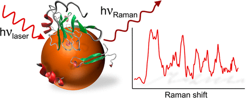
Surface-enhanced Raman scattering (SERS) can provide information on the structure, composition, and interaction of molecules in the proximity of gold nanoparticles, thereby enabling studies of adsorbed biomolecules in vivo. Here, the processing of the protein corona and the corresponding protein–nanoparticle interactions in live J774 cells incubated with gold nanoparticles was characterized by SERS. Samples of isolated cytoplasm, devoid of active processing, of the same cell line were used as references. The occurrence of the most important SERS signals was compared in both types of samples. The comparison of signal abundances, supported by multivariate assessment, suggests a decreased nanoparticle–peptide backbone interaction and an increased contribution of denatured proteins in endolysosomal compartments, indicating an interaction of protein fragments with the gold nanoparticles in the endolysosome of the living cells. To study the protein fragmentation in a model and to confirm the assignment of specific spectral signatures in the live cell spectra, SERS data were collected from a solution of bovine serum albumin (BSA) digested by trypsin as an enzymatic model and from solutions of intact BSA and trypsin. The spectra from the enzymatic model confirm the strong interaction of protein fragments with the gold nanoparticles in the endolysosomal compartments. By proteomic analysis, using combined sodium dodecyl sulfate-polyacrylamide gel electrophoresis and high-performance liquid chromatography–electrospray ionization-tandem mass spectrometry of the extracted hard corona, we directly identified protein fragments, some originating from the culture medium. The results illustrate the use of appropriate models for the validation of SERS spectra and have potential implications for further developments of SERS as an in vivo analytical and biomedical tool.
中文翻译:

在活细胞表面增强拉曼散射中观察到的金纳米粒子电晕中蛋白质的断裂。
表面增强拉曼散射(SERS)可以提供有关金纳米颗粒附近分子的结构,组成和相互作用的信息,从而可以研究体内吸附的生物分子。在这里,用SERS表征了在与金纳米颗粒一起孵育的活J774细胞中蛋白质电晕的加工以及相应的蛋白质-纳米颗粒相互作用。将没有活性处理的相同细胞系的分离的细胞质样品用作参考。比较了两种样品中最重要的SERS信号的发生情况。信号丰度的比较得到多变量评估的支持,表明减少的纳米颗粒-肽主链相互作用和变性的蛋白质在溶酶体区室中的贡献增加,表明蛋白质片段与活细胞的溶酶体中的金纳米颗粒相互作用。要研究模型中的蛋白质碎片并确认活细胞光谱中特定光谱特征的分配,SERS数据是从被胰蛋白酶消化的牛血清白蛋白(BSA)溶液(作为酶模型)以及完整的BSA和胰蛋白酶的溶液中收集的。酶模型的光谱证实了溶酶体区室中蛋白质片段与金纳米颗粒之间的强相互作用。通过蛋白质组学分析,结合十二烷基硫酸钠-聚丙烯酰胺凝胶电泳和高效液相色谱-电喷雾电离串联质谱法对提取的硬质电晕进行分离,我们直接鉴定了蛋白质片段,其中一些源自培养基。结果表明,使用适当的模型来验证SERS光谱,对于SERS的进一步开发具有潜在的潜在意义。体内分析和生物医学工具。
更新日期:2020-05-18
中文翻译:

在活细胞表面增强拉曼散射中观察到的金纳米粒子电晕中蛋白质的断裂。
表面增强拉曼散射(SERS)可以提供有关金纳米颗粒附近分子的结构,组成和相互作用的信息,从而可以研究体内吸附的生物分子。在这里,用SERS表征了在与金纳米颗粒一起孵育的活J774细胞中蛋白质电晕的加工以及相应的蛋白质-纳米颗粒相互作用。将没有活性处理的相同细胞系的分离的细胞质样品用作参考。比较了两种样品中最重要的SERS信号的发生情况。信号丰度的比较得到多变量评估的支持,表明减少的纳米颗粒-肽主链相互作用和变性的蛋白质在溶酶体区室中的贡献增加,表明蛋白质片段与活细胞的溶酶体中的金纳米颗粒相互作用。要研究模型中的蛋白质碎片并确认活细胞光谱中特定光谱特征的分配,SERS数据是从被胰蛋白酶消化的牛血清白蛋白(BSA)溶液(作为酶模型)以及完整的BSA和胰蛋白酶的溶液中收集的。酶模型的光谱证实了溶酶体区室中蛋白质片段与金纳米颗粒之间的强相互作用。通过蛋白质组学分析,结合十二烷基硫酸钠-聚丙烯酰胺凝胶电泳和高效液相色谱-电喷雾电离串联质谱法对提取的硬质电晕进行分离,我们直接鉴定了蛋白质片段,其中一些源自培养基。结果表明,使用适当的模型来验证SERS光谱,对于SERS的进一步开发具有潜在的潜在意义。体内分析和生物医学工具。


 京公网安备 11010802027423号