当前位置:
X-MOL 学术
›
Nat. Commun.
›
论文详情
Our official English website, www.x-mol.net, welcomes your
feedback! (Note: you will need to create a separate account there.)
Real-time non-contact cellular imaging and angiography of human cornea and limbus with common-path full-field/SD OCT.
Nature Communications ( IF 14.7 ) Pub Date : 2020-04-20 , DOI: 10.1038/s41467-020-15792-x Viacheslav Mazlin 1 , Peng Xiao 1, 2 , Jules Scholler 1 , Kristina Irsch 3, 4 , Kate Grieve 3, 4 , Mathias Fink 1 , A Claude Boccara 1
Nature Communications ( IF 14.7 ) Pub Date : 2020-04-20 , DOI: 10.1038/s41467-020-15792-x Viacheslav Mazlin 1 , Peng Xiao 1, 2 , Jules Scholler 1 , Kristina Irsch 3, 4 , Kate Grieve 3, 4 , Mathias Fink 1 , A Claude Boccara 1
Affiliation

|
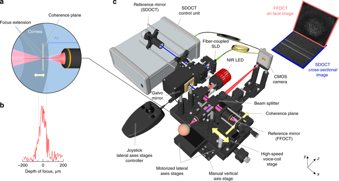
In today's clinics, a cell-resolution view of the cornea can be achieved only with a confocal microscope (IVCM) in contact with the eye. Here, we present a common-path full-field/spectral-domain OCT microscope (FF/SD OCT), which enables cell-detail imaging of the entire ocular surface in humans (central and peripheral cornea, limbus, sclera, tear film) without contact and in real-time. Real-time performance is achieved through rapid axial eye tracking and simultaneous defocusing correction. Images contain cells and nerves, which can be quantified over a millimetric field-of-view, beyond the capability of IVCM and conventional OCT. In the limbus, palisades of Vogt, vessels, and blood flow can be resolved with high contrast without contrast agent injection. The fast imaging speed of 275 frames/s (0.6 billion pixels/s) allows direct monitoring of blood flow dynamics, enabling creation of high-resolution velocity maps. Tear flow velocity and evaporation time can be measured without fluorescein administration.
中文翻译:

使用共路径全视野/SD OCT 对人类角膜和角膜缘进行实时非接触式细胞成像和血管造影。
在当今的诊所中,只有使用与眼睛接触的共焦显微镜 (IVCM) 才能获得角膜的细胞分辨率视图。在这里,我们提出了一种共光路全视野/谱域 OCT 显微镜 (FF/SD OCT),它能够对人类整个眼表(中央和周边角膜、角膜缘、巩膜、泪膜)进行细胞细节成像无接触且实时。通过快速轴眼跟踪和同时散焦校正来实现实时性能。图像包含细胞和神经,可以在毫米级视野内进行量化,超出了 IVCM 和传统 OCT 的能力。在角膜缘,Vogt栅栏、血管和血流可以通过高对比度分辨出来,而无需注射造影剂。 275 帧/秒(6 亿像素/秒)的快速成像速度可以直接监测血流动力学,从而创建高分辨率速度图。无需注射荧光素即可测量泪液流速和蒸发时间。
更新日期:2020-04-24
中文翻译:

使用共路径全视野/SD OCT 对人类角膜和角膜缘进行实时非接触式细胞成像和血管造影。
在当今的诊所中,只有使用与眼睛接触的共焦显微镜 (IVCM) 才能获得角膜的细胞分辨率视图。在这里,我们提出了一种共光路全视野/谱域 OCT 显微镜 (FF/SD OCT),它能够对人类整个眼表(中央和周边角膜、角膜缘、巩膜、泪膜)进行细胞细节成像无接触且实时。通过快速轴眼跟踪和同时散焦校正来实现实时性能。图像包含细胞和神经,可以在毫米级视野内进行量化,超出了 IVCM 和传统 OCT 的能力。在角膜缘,Vogt栅栏、血管和血流可以通过高对比度分辨出来,而无需注射造影剂。 275 帧/秒(6 亿像素/秒)的快速成像速度可以直接监测血流动力学,从而创建高分辨率速度图。无需注射荧光素即可测量泪液流速和蒸发时间。


 京公网安备 11010802027423号