当前位置:
X-MOL 学术
›
Nat. Commun.
›
论文详情
Our official English website, www.x-mol.net, welcomes your
feedback! (Note: you will need to create a separate account there.)
Computational analysis of pathological images enables a better diagnosis of TFE3 Xp11.2 translocation renal cell carcinoma.
Nature Communications ( IF 14.7 ) Pub Date : 2020-04-14 , DOI: 10.1038/s41467-020-15671-5
Jun Cheng 1 , Zhi Han 2, 3 , Rohit Mehra 4 , Wei Shao 2 , Michael Cheng 2 , Qianjin Feng 5 , Dong Ni 1 , Kun Huang 2, 3 , Liang Cheng 6 , Jie Zhang 7
Nature Communications ( IF 14.7 ) Pub Date : 2020-04-14 , DOI: 10.1038/s41467-020-15671-5
Jun Cheng 1 , Zhi Han 2, 3 , Rohit Mehra 4 , Wei Shao 2 , Michael Cheng 2 , Qianjin Feng 5 , Dong Ni 1 , Kun Huang 2, 3 , Liang Cheng 6 , Jie Zhang 7
Affiliation
|
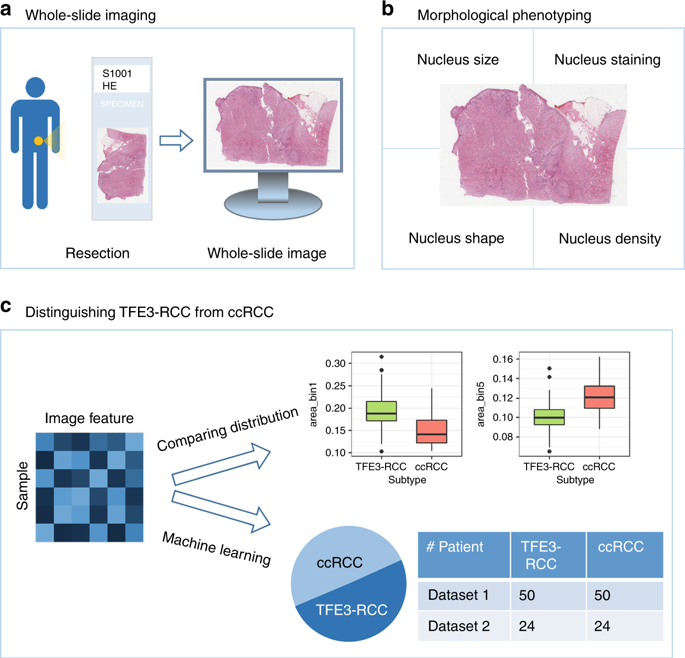
TFE3 Xp11.2 translocation renal cell carcinoma (TFE3-RCC) generally progresses more aggressively compared with other RCC subtypes, but it is challenging to diagnose TFE3-RCC by traditional visual inspection of pathological images. In this study, we collect hematoxylin and eosin- stained histopathology whole-slide images of 74 TFE3-RCC cases (the largest cohort to date) and 74 clear cell RCC cases (ccRCC, the most common RCC subtype) with matched gender and tumor grade. An automatic computational pipeline is implemented to extract image features. Comparative study identifies 52 image features with significant differences between TFE3-RCC and ccRCC. Machine learning models are built to distinguish TFE3-RCC from ccRCC. Tests of the classification models on an external validation set reveal high accuracy with areas under ROC curve ranging from 0.842 to 0.894. Our results suggest that automatically derived image features can capture subtle morphological differences between TFE3-RCC and ccRCC and contribute to a potential guideline for TFE3-RCC diagnosis.
中文翻译:

病理图像的计算分析可以更好地诊断TFE3 Xp11.2易位性肾细胞癌。
与其他RCC亚型相比,TFE3 Xp11.2易位性肾细胞癌(TFE3-RCC)的进展通常更为积极,但是通过传统的病理影像学检查来诊断TFE3-RCC颇具挑战性。在这项研究中,我们收集了74例TFE3-RCC病例(迄今为止最大的队列)和74例透明细胞RCC病例(ccRCC,最常见的RCC亚型)的苏木精和曙红染色的组织病理学全片图像,这些性别和肿瘤等级相匹配。实现了自动计算流水线以提取图像特征。比较研究确定了52个图像特征,TFE3-RCC和ccRCC之间存在显着差异。建立了机器学习模型以区分TFE3-RCC和ccRCC。在外部验证集上对分类模型进行的测试表明,ROC曲线下的面积为0,具有很高的准确性。842至0.894。我们的结果表明,自动导出的图像特征可以捕获TFE3-RCC和ccRCC之间的细微形态差异,并有助于TFE3-RCC诊断的潜在指南。
更新日期:2020-04-24
中文翻译:
病理图像的计算分析可以更好地诊断TFE3 Xp11.2易位性肾细胞癌。
与其他RCC亚型相比,TFE3 Xp11.2易位性肾细胞癌(TFE3-RCC)的进展通常更为积极,但是通过传统的病理影像学检查来诊断TFE3-RCC颇具挑战性。在这项研究中,我们收集了74例TFE3-RCC病例(迄今为止最大的队列)和74例透明细胞RCC病例(ccRCC,最常见的RCC亚型)的苏木精和曙红染色的组织病理学全片图像,这些性别和肿瘤等级相匹配。实现了自动计算流水线以提取图像特征。比较研究确定了52个图像特征,TFE3-RCC和ccRCC之间存在显着差异。建立了机器学习模型以区分TFE3-RCC和ccRCC。在外部验证集上对分类模型进行的测试表明,ROC曲线下的面积为0,具有很高的准确性。842至0.894。我们的结果表明,自动导出的图像特征可以捕获TFE3-RCC和ccRCC之间的细微形态差异,并有助于TFE3-RCC诊断的潜在指南。


 京公网安备 11010802027423号