当前位置:
X-MOL 学术
›
J. Invertebr. Pathol.
›
论文详情
Our official English website, www.x-mol.net, welcomes your
feedback! (Note: you will need to create a separate account there.)
In situ hybridization revealed wide distribution of Haliotid herpesvirus 1 in infected small abalone, Haliotis diversicolor supertexta.
Journal of Invertebrate Pathology ( IF 3.6 ) Pub Date : 2020-03-19 , DOI: 10.1016/j.jip.2020.107356 Chang-Ming Bai , Ya-Nan Li , Pen-Heng Chang , Jing-Zhe Jiang , Lu-Sheng Xin , Chen Li , Jiang-Yong Wang , Chong-Ming Wang
Journal of Invertebrate Pathology ( IF 3.6 ) Pub Date : 2020-03-19 , DOI: 10.1016/j.jip.2020.107356 Chang-Ming Bai , Ya-Nan Li , Pen-Heng Chang , Jing-Zhe Jiang , Lu-Sheng Xin , Chen Li , Jiang-Yong Wang , Chong-Ming Wang

|
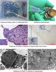
Ganglioneuritis was the primary pathologic change in infected abalone associated with Haliotid herpesvirus 1 (HaHV-1) infection, which eventually became known as abalone viral ganglioneuritis (AVG). However, the distribution of HaHV-1 in the other tissues and organs of infected abalone has not been systemically investigated. In the present study, the distribution of HaHV-1-CN2003 variant in different organs of small abalone, Haliotis diversicolor supertexta, collected at seven different time points post experimental infection, was investigated with histopathological examination and in situ hybridization (ISH) of HaHV-1 DNA. ISH signals were first observed in pedal ganglia at 48 h post injection, and were consistently observed in this tissue of challenged abalone. At the same time, increased cellularity accompanied by ISH signals was observed in some peripheral ganglia of mantle and kidney. At the end of infection period, lesions and co-localized ISH signals in infiltrated cells were detected occasionally in the mantle and hepatopancreas. Transmission electron microscope analysis revealed the presence of herpes-like viral particles in haemocyte nuclei of infected abalone. Our results indicated that, although HaHV-1-CN2003 was primarily neurotropic, it could infect other tissues including haemocytes.
中文翻译:

原位杂交显示在感染的小鲍鱼Haliotis diversicolor supertexta中,Haliotid疱疹病毒1广泛分布。
神经节神经炎是与鲍状疱疹病毒1(HaHV-1)感染相关的被感染鲍鱼的主要病理变化,最终被称为鲍鱼病毒性神经节神经炎(AVG)。但是,HaHV-1在受感染的鲍鱼的其他组织和器官中的分布尚未得到系统的研究。在本研究中,通过组织病理学检查和原位杂交(ISH)研究了在实验感染后七个不同时间点采集的HaHV-1-CN2003变体在小鲍鲍鱼不同器官中的分布。 1个DNA。ISH信号首先在注射后48小时在踏板神经节中观察到,并在该受攻击的鲍鱼组织中一致观察到。同时,在地幔和肾脏的一些外周神经节中观察到细胞增多并伴有ISH信号。在感染期结束时,偶尔会在地幔和肝胰腺中检测到浸润细胞中的病变和共定位的ISH信号。透射电子显微镜分析显示在感染的鲍鱼血细胞核中存在疱疹样病毒颗粒。我们的结果表明,尽管HaHV-1-CN2003主要是神经质的,但它可以感染其他组织,包括血细胞。
更新日期:2020-04-21
中文翻译:

原位杂交显示在感染的小鲍鱼Haliotis diversicolor supertexta中,Haliotid疱疹病毒1广泛分布。
神经节神经炎是与鲍状疱疹病毒1(HaHV-1)感染相关的被感染鲍鱼的主要病理变化,最终被称为鲍鱼病毒性神经节神经炎(AVG)。但是,HaHV-1在受感染的鲍鱼的其他组织和器官中的分布尚未得到系统的研究。在本研究中,通过组织病理学检查和原位杂交(ISH)研究了在实验感染后七个不同时间点采集的HaHV-1-CN2003变体在小鲍鲍鱼不同器官中的分布。 1个DNA。ISH信号首先在注射后48小时在踏板神经节中观察到,并在该受攻击的鲍鱼组织中一致观察到。同时,在地幔和肾脏的一些外周神经节中观察到细胞增多并伴有ISH信号。在感染期结束时,偶尔会在地幔和肝胰腺中检测到浸润细胞中的病变和共定位的ISH信号。透射电子显微镜分析显示在感染的鲍鱼血细胞核中存在疱疹样病毒颗粒。我们的结果表明,尽管HaHV-1-CN2003主要是神经质的,但它可以感染其他组织,包括血细胞。


 京公网安备 11010802027423号