当前位置:
X-MOL 学术
›
Spectrochim. Acta. A Mol. Biomol. Spectrosc.
›
论文详情
Our official English website, www.x-mol.net, welcomes your
feedback! (Note: you will need to create a separate account there.)
SERS studies on normal epithelial and cancer cells derived from clinical breast cancer specimens.
Spectrochimica Acta Part A: Molecular and Biomolecular Spectroscopy ( IF 4.3 ) Pub Date : 2020-04-12 , DOI: 10.1016/j.saa.2020.118364 LiShengNan Shen 1 , Ye Du 1 , Na Wei 2 , Qian Li 1 , SiMin Li 1 , TianMeng Sun 3 , Shuping Xu 4 , Han Wang 5 , XiaXia Man 6 , Bing Han 1
Spectrochimica Acta Part A: Molecular and Biomolecular Spectroscopy ( IF 4.3 ) Pub Date : 2020-04-12 , DOI: 10.1016/j.saa.2020.118364 LiShengNan Shen 1 , Ye Du 1 , Na Wei 2 , Qian Li 1 , SiMin Li 1 , TianMeng Sun 3 , Shuping Xu 4 , Han Wang 5 , XiaXia Man 6 , Bing Han 1
Affiliation

|
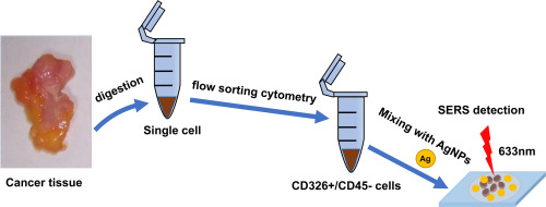
Surface-enhanced Raman scattering (SERS) spectroscopy of single-cell suspensions obtained from fresh specimens of breast cancer tissue and normal breast tissue by mechanical enzymatic digestion was obtained and analysed, which is different from most Raman studies using breast cancer cell lines. Random forest classification was implemented to develop effective diagnostic algorithms for the classification of SERS of different typed cells. We first examined the SERS spectra of the primary breast cancer single cell and normal epithelial single cell obtained by flow sorting cytometry due to their biomarkers of CD326+/CD45-. Comparison analyses on their SERS spectra disclose that the nucleic acid and protein levels of the primary breast cancer single cell are higher than those of the normal epithelial single cell, while the lipids are at a relatively lower level. An important finding is that the cholesterol, palmitic acid, and sphingomyelin in the cancer cell profiles exhibit stronger than those of normal cells, while the glycans are at a relatively lower level. Furthermore, the standard deviation (SD) of the normal epithelial single cell is larger than that of the breast cancer cell, and the SD of the primary breast cancer single cell is more obvious than that of the normal epithelial cells. In addition, the prospective application of an algorithm to the dataset results in an accuracy of 78.2%, a precision of 75.5%, and a recall of 66.7%. The breast cancer diagnostic model laid a solid foundation for judgment of breast-conserving surgical margins and early diagnosis of breast cancer.
中文翻译:

SERS研究源自临床乳腺癌样本的正常上皮和癌细胞。
获得并分析了通过机械酶消化从乳腺癌组织和正常乳腺组织的新鲜标本中获得的单细胞悬液的表面增强拉曼散射(SERS)光谱,这与大多数使用乳腺癌细胞系的拉曼研究不同。实施随机森林分类以开发有效的诊断算法,用于分类不同类型细胞的SERS。我们首先检查了通过流式细胞术获得的原发性乳腺癌单细胞和正常上皮单细胞的SERS光谱,这是由于它们具有CD326 + / CD45-的生物标记。对他们的SERS光谱进行的比较分析表明,原发性乳腺癌单细胞的核酸和蛋白质水平高于正常上皮单细胞的核酸和蛋白质水平,而脂质则相对较低。一个重要发现是,癌细胞谱中的胆固醇,棕榈酸和鞘磷脂比普通细胞表现出更强的功能,而聚糖的水平相对较低。此外,正常上皮单细胞的标准差(SD)大于乳腺癌细胞的标准差,并且原发性乳腺癌单细胞的SD比正常上皮细胞的SD更明显。此外,将算法应用于数据集的预期准确性为78.2%,准确性为75.5%,召回率为66.7%。乳腺癌诊断模型为判断保留乳腺癌的手术切缘和早期诊断奠定了坚实的基础。鞘磷脂和鞘磷脂在癌细胞中的表现要强于正常细胞,而聚糖含量则相对较低。此外,正常上皮单细胞的标准差(SD)大于乳腺癌细胞的标准差,并且原发性乳腺癌单细胞的SD比正常上皮细胞的SD更明显。此外,将算法应用于数据集的预期准确性为78.2%,准确性为75.5%,召回率为66.7%。乳腺癌诊断模型为判断保留乳腺癌的手术切缘和早期诊断奠定了坚实的基础。鞘磷脂和鞘磷脂在癌细胞中的表现要强于正常细胞,而聚糖含量则相对较低。此外,正常上皮单细胞的标准差(SD)大于乳腺癌细胞的标准差,并且原发性乳腺癌单细胞的SD比正常上皮细胞的SD更明显。此外,将算法应用于数据集的预期准确性为78.2%,准确性为75.5%,召回率为66.7%。乳腺癌诊断模型为判断保留乳腺癌的手术切缘和早期诊断奠定了坚实的基础。正常上皮单细胞的标准偏差(SD)比乳腺癌细胞大,而原发性乳腺癌单细胞的SD比正常上皮细胞更明显。此外,将算法应用于数据集的预期准确性为78.2%,准确性为75.5%,召回率为66.7%。乳腺癌诊断模型为判断保留乳腺癌的手术切缘和早期诊断奠定了坚实的基础。正常上皮单细胞的标准偏差(SD)比乳腺癌细胞大,而原发性乳腺癌单细胞的SD比正常上皮细胞更明显。此外,将算法应用于数据集的预期准确性为78.2%,准确性为75.5%,召回率为66.7%。乳腺癌诊断模型为判断保留乳腺癌的手术切缘和早期诊断奠定了坚实的基础。
更新日期:2020-04-13
中文翻译:

SERS研究源自临床乳腺癌样本的正常上皮和癌细胞。
获得并分析了通过机械酶消化从乳腺癌组织和正常乳腺组织的新鲜标本中获得的单细胞悬液的表面增强拉曼散射(SERS)光谱,这与大多数使用乳腺癌细胞系的拉曼研究不同。实施随机森林分类以开发有效的诊断算法,用于分类不同类型细胞的SERS。我们首先检查了通过流式细胞术获得的原发性乳腺癌单细胞和正常上皮单细胞的SERS光谱,这是由于它们具有CD326 + / CD45-的生物标记。对他们的SERS光谱进行的比较分析表明,原发性乳腺癌单细胞的核酸和蛋白质水平高于正常上皮单细胞的核酸和蛋白质水平,而脂质则相对较低。一个重要发现是,癌细胞谱中的胆固醇,棕榈酸和鞘磷脂比普通细胞表现出更强的功能,而聚糖的水平相对较低。此外,正常上皮单细胞的标准差(SD)大于乳腺癌细胞的标准差,并且原发性乳腺癌单细胞的SD比正常上皮细胞的SD更明显。此外,将算法应用于数据集的预期准确性为78.2%,准确性为75.5%,召回率为66.7%。乳腺癌诊断模型为判断保留乳腺癌的手术切缘和早期诊断奠定了坚实的基础。鞘磷脂和鞘磷脂在癌细胞中的表现要强于正常细胞,而聚糖含量则相对较低。此外,正常上皮单细胞的标准差(SD)大于乳腺癌细胞的标准差,并且原发性乳腺癌单细胞的SD比正常上皮细胞的SD更明显。此外,将算法应用于数据集的预期准确性为78.2%,准确性为75.5%,召回率为66.7%。乳腺癌诊断模型为判断保留乳腺癌的手术切缘和早期诊断奠定了坚实的基础。鞘磷脂和鞘磷脂在癌细胞中的表现要强于正常细胞,而聚糖含量则相对较低。此外,正常上皮单细胞的标准差(SD)大于乳腺癌细胞的标准差,并且原发性乳腺癌单细胞的SD比正常上皮细胞的SD更明显。此外,将算法应用于数据集的预期准确性为78.2%,准确性为75.5%,召回率为66.7%。乳腺癌诊断模型为判断保留乳腺癌的手术切缘和早期诊断奠定了坚实的基础。正常上皮单细胞的标准偏差(SD)比乳腺癌细胞大,而原发性乳腺癌单细胞的SD比正常上皮细胞更明显。此外,将算法应用于数据集的预期准确性为78.2%,准确性为75.5%,召回率为66.7%。乳腺癌诊断模型为判断保留乳腺癌的手术切缘和早期诊断奠定了坚实的基础。正常上皮单细胞的标准偏差(SD)比乳腺癌细胞大,而原发性乳腺癌单细胞的SD比正常上皮细胞更明显。此外,将算法应用于数据集的预期准确性为78.2%,准确性为75.5%,召回率为66.7%。乳腺癌诊断模型为判断保留乳腺癌的手术切缘和早期诊断奠定了坚实的基础。


 京公网安备 11010802027423号