当前位置:
X-MOL 学术
›
Macromol. Biosci.
›
论文详情
Our official English website, www.x-mol.net, welcomes your
feedback! (Note: you will need to create a separate account there.)
Polycaprolactone Electrospun Fiber Mats Prepared Using Benign Solvents: Blending with Copper(II)-Chitosan Increases the Secretion of Vascular Endothelial Growth Factor in a Bone Marrow Stromal Cell Line.
Macromolecular Bioscience ( IF 4.4 ) Pub Date : 2020-02-05 , DOI: 10.1002/mabi.201900355 Lukas Gritsch 1, 2 , Liliana Liverani 1 , Christopher Lovell 2 , Aldo R Boccaccini 1
Macromolecular Bioscience ( IF 4.4 ) Pub Date : 2020-02-05 , DOI: 10.1002/mabi.201900355 Lukas Gritsch 1, 2 , Liliana Liverani 1 , Christopher Lovell 2 , Aldo R Boccaccini 1
Affiliation

|
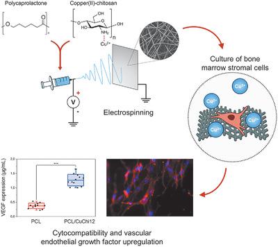
Inducing the formation of new blood vessels (angiogenesis) is an essential requirement for successful tissue engineering. Approaches have been proposed to enhance angiogenesis using growth factors and other biomolecules; however, this approaches present drawbacks in terms of high cost and patient safety. Copper is known to effectively regulate angiogenesis and can offer a more cost‐effective alternative than the direct use of growth factors. With this study, a strategy to incorporate copper in electrospun fibrous scaffolds with pro‐angiogenic properties is presented. Polycaprolactone (PCL) and copper(II)‐chitosan are electrospun using benign solvents. The morphological and physicochemical properties of the fiber mats are investigated through scanning electron microscopy (SEM), static contact angle measurements, energy dispersive X‐ray, and Fourier‐transform infrared spectroscopies. Scaffold stability in phosphate buffered saline at 37 °C is monitored over 1 week. A bone marrow stromal cell line (ST‐2) is cultured for 7 days and its behavior is evaluated using SEM, fluorescence microscopy and a tetrazolium salt‐based colorimetric assay. Results confirm that PCL/copper(II)‐chitosan is suitable for electrospinning. The fiber mats are biocompatible and favor cell colonization and infiltration. Most notably, the angiogenic potential of PCL/copper(II)‐chitosan blends is confirmed by a three‐fold increase in VEGF secretion by ST‐2 cells in the presence of copper(II)‐chitosan.
中文翻译:

使用良性溶剂制备的聚己内酯电纺纤维毡:与铜(II)-壳聚糖共混可增加骨髓基质细胞系中血管内皮生长因子的分泌。
诱导新血管的形成(血管生成)是成功进行组织工程的基本要求。已经提出了使用生长因子和其他生物分子增强血管生成的方法。然而,这种方法在高成本和患者安全方面存在缺点。众所周知,铜可以有效地调节血管生成,与直接使用生长因子相比,铜可以提供更具成本效益的替代方法。通过这项研究,提出了一种将铜结合到具有促血管生成特性的电纺纤维支架中的策略。使用良性溶剂对聚己内酯(PCL)和铜(II)-壳聚糖进行静电纺丝。通过扫描电子显微镜(SEM),静态接触角测量,能量色散X射线研究纤维毡的形态和理化性质 和傅立叶变换红外光谱。在1周内监测在37°C的磷酸盐缓冲盐水中的支架稳定性。将骨髓基质细胞系(ST-2)培养7天,并使用SEM,荧光显微镜和基于四唑鎓盐的比色分析评估其行为。结果证实PCL /铜(II)-壳聚糖适合静电纺丝。纤维垫具有生物相容性,有利于细胞定植和浸润。最值得注意的是,在铜(II)-壳聚糖存在下,ST-2细胞的VEGF分泌增加了三倍,从而证实了PCL /铜(II)-壳聚糖混合物的血管生成潜力。将骨髓基质细胞系(ST-2)培养7天,并使用SEM,荧光显微镜和基于四唑鎓盐的比色分析评估其行为。结果证实PCL /铜(II)-壳聚糖适合静电纺丝。纤维垫具有生物相容性,有利于细胞定植和浸润。最值得注意的是,在铜(II)-壳聚糖存在下,ST-2细胞的VEGF分泌增加了三倍,从而证实了PCL /铜(II)-壳聚糖混合物的血管生成潜力。将骨髓基质细胞系(ST-2)培养7天,并使用SEM,荧光显微镜和基于四唑鎓盐的比色分析评估其行为。结果证实PCL /铜(II)-壳聚糖适合静电纺丝。纤维垫具有生物相容性,有利于细胞定植和浸润。最值得注意的是,在铜(II)-壳聚糖存在下,ST-2细胞的VEGF分泌增加了三倍,从而证实了PCL /铜(II)-壳聚糖混合物的血管生成潜力。
更新日期:2020-02-05
中文翻译:

使用良性溶剂制备的聚己内酯电纺纤维毡:与铜(II)-壳聚糖共混可增加骨髓基质细胞系中血管内皮生长因子的分泌。
诱导新血管的形成(血管生成)是成功进行组织工程的基本要求。已经提出了使用生长因子和其他生物分子增强血管生成的方法。然而,这种方法在高成本和患者安全方面存在缺点。众所周知,铜可以有效地调节血管生成,与直接使用生长因子相比,铜可以提供更具成本效益的替代方法。通过这项研究,提出了一种将铜结合到具有促血管生成特性的电纺纤维支架中的策略。使用良性溶剂对聚己内酯(PCL)和铜(II)-壳聚糖进行静电纺丝。通过扫描电子显微镜(SEM),静态接触角测量,能量色散X射线研究纤维毡的形态和理化性质 和傅立叶变换红外光谱。在1周内监测在37°C的磷酸盐缓冲盐水中的支架稳定性。将骨髓基质细胞系(ST-2)培养7天,并使用SEM,荧光显微镜和基于四唑鎓盐的比色分析评估其行为。结果证实PCL /铜(II)-壳聚糖适合静电纺丝。纤维垫具有生物相容性,有利于细胞定植和浸润。最值得注意的是,在铜(II)-壳聚糖存在下,ST-2细胞的VEGF分泌增加了三倍,从而证实了PCL /铜(II)-壳聚糖混合物的血管生成潜力。将骨髓基质细胞系(ST-2)培养7天,并使用SEM,荧光显微镜和基于四唑鎓盐的比色分析评估其行为。结果证实PCL /铜(II)-壳聚糖适合静电纺丝。纤维垫具有生物相容性,有利于细胞定植和浸润。最值得注意的是,在铜(II)-壳聚糖存在下,ST-2细胞的VEGF分泌增加了三倍,从而证实了PCL /铜(II)-壳聚糖混合物的血管生成潜力。将骨髓基质细胞系(ST-2)培养7天,并使用SEM,荧光显微镜和基于四唑鎓盐的比色分析评估其行为。结果证实PCL /铜(II)-壳聚糖适合静电纺丝。纤维垫具有生物相容性,有利于细胞定植和浸润。最值得注意的是,在铜(II)-壳聚糖存在下,ST-2细胞的VEGF分泌增加了三倍,从而证实了PCL /铜(II)-壳聚糖混合物的血管生成潜力。


 京公网安备 11010802027423号