Our official English website, www.x-mol.net, welcomes your
feedback! (Note: you will need to create a separate account there.)
The upper cervical spinal cord in ALS assessed by cross-sectional and longitudinal 3T MRI.
Scientific Reports ( IF 3.8 ) Pub Date : 2020-02-04 , DOI: 10.1038/s41598-020-58687-z
Thomas Wimmer 1 , Frank Schreiber 1, 2 , Nathalie Hensiek 1 , Cornelia Garz 1, 2, 3 , Jörn Kaufmann 1 , Judith Machts 1, 2 , Susanne Vogt 1 , Johannes Prudlo 4, 5 , Reinhard Dengler 6 , Susanne Petri 6 , Hans-Jochen Heinze 1, 2, 3, 7 , Peter J Nestor 8, 9 , Stefan Vielhaber 1, 2, 7 , Stefanie Schreiber 1, 2, 7
Affiliation
|
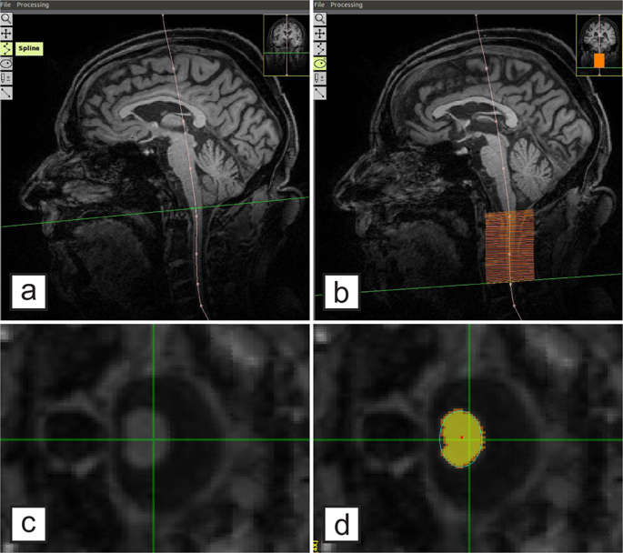
The upper cervical spinal cord is measured in a large longitudinal amyotrophic lateral sclerosis (ALS) cohort to evaluate its role as a biomarker. Specifically, the cervical spinal cord´s cross-sectional area (CSA) in plane of the segments C1-C3 was measured semi-automatically with T1-weighted 3T MRI sequences in 158 ALS patients and 86 controls. Six-month longitudinal follow-up MRI scans were analyzed in 103 patients. Compared to controls, in ALS there was a significant mean spinal cord atrophy (63.8 mm² vs. 60.8 mm², p = 0.001) which showed a trend towards worsening over time (mean spinal cord CSA decrease from 61.4 mm² to 60.6 mm² after 6 months, p = 0.06). Findings were most pronounced in the caudal segments of the upper cervical spinal cord and in limb-onset ALS. Baseline CSA was related to the revised ALS functional rating scale, disease duration, precentral gyrus thickness and total brain gray matter volume. In conclusion, spinal cord atrophy as assessed in brain MRIs in ALS patients mirrors the extent of overall neurodegeneration and parallels disease severity.
中文翻译:

通过横截面和纵向3T MRI评估ALS中的上颈脊髓。
在大型纵向肌萎缩性侧索硬化症(ALS)队列中测量上颈脊髓,以评估其作为生物标志物的作用。具体而言,使用T1加权3T MRI序列半自动测量了158名ALS患者和86名对照的C1-C3段平面中的颈脊髓横截面积(CSA)。对103例患者进行了为期六个月的纵向随访MRI扫描。与对照组相比,ALS患者的平均脊髓萎缩显着(63.8mm²对60.8mm²,p = 0.001),并且随着时间的推移呈恶化趋势(6个月后,平均脊髓CSA从61.4mm²降至60.6mm², p = 0.06)。在上颈脊髓的尾段和四肢发作的ALS中发现最明显。基准CSA与修订后的ALS功能评分表相关,疾病持续时间,中央前回厚度和总脑灰质体积。总之,在ALS患者的脑部MRI中评估的脊髓萎缩反映了总体神经退行性变的程度和疾病严重程度的相似性。
更新日期:2020-02-04
中文翻译:
通过横截面和纵向3T MRI评估ALS中的上颈脊髓。
在大型纵向肌萎缩性侧索硬化症(ALS)队列中测量上颈脊髓,以评估其作为生物标志物的作用。具体而言,使用T1加权3T MRI序列半自动测量了158名ALS患者和86名对照的C1-C3段平面中的颈脊髓横截面积(CSA)。对103例患者进行了为期六个月的纵向随访MRI扫描。与对照组相比,ALS患者的平均脊髓萎缩显着(63.8mm²对60.8mm²,p = 0.001),并且随着时间的推移呈恶化趋势(6个月后,平均脊髓CSA从61.4mm²降至60.6mm², p = 0.06)。在上颈脊髓的尾段和四肢发作的ALS中发现最明显。基准CSA与修订后的ALS功能评分表相关,疾病持续时间,中央前回厚度和总脑灰质体积。总之,在ALS患者的脑部MRI中评估的脊髓萎缩反映了总体神经退行性变的程度和疾病严重程度的相似性。


 京公网安备 11010802027423号