Cellular & Molecular Immunology ( IF 21.8 ) Pub Date : 2020-01-27 , DOI: 10.1038/s41423-019-0352-8
Eirini Nikolouli 1 , Yassin Elfaki 1 , Susanne Herppich 1 , Carsten Schelmbauer 2 , Michael Delacher 3 , Christine Falk 4 , Ilgiz A Mufazalov 2 , Ari Waisman 2 , Markus Feuerer 3 , Jochen Huehn 1, 5
|
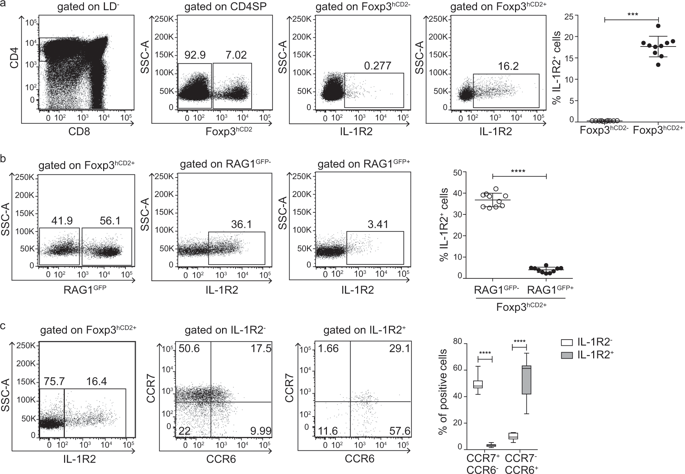
The vast majority of Foxp3+ regulatory T cells (Tregs) are generated in the thymus, and several factors, such as cytokines and unique thymic antigen-presenting cells, are known to contribute to the development of these thymus-derived Tregs (tTregs). Here, we report the existence of a specific subset of Foxp3+ Tregs within the thymus that is characterized by the expression of IL-1R2, which is a decoy receptor for the inflammatory cytokine IL-1. Detailed flow cytometric analysis of the thymocytes from Foxp3hCD2xRAG1GFP reporter mice revealed that the IL-1R2+ Tregs are mainly RAG1GFP– and CCR6+CCR7–, demonstrating that these Tregs are recirculating cells entering the thymus from the periphery and that they have an activated phenotype. In the spleen, the majority of IL-1R2+ Tregs express neuropilin-1 (Nrp-1) and Helios, suggesting a thymic origin for these Tregs. Interestingly, among all tissues studied, the highest frequency of IL-1R2+ Tregs was observed in the thymus, indicating preferential recruitment of this Treg subset by the thymus. Using fetal thymic organ cultures (FTOCs), we demonstrated that increased concentrations of exogenous IL-1β blocked intrathymic Treg development, resulting in a decreased frequency of CD25+Foxp3+ tTregs and an accumulation of CD25+Foxp3− Treg precursors. Interestingly, the addition of IL-1R2+ Tregs, but not IL-1R2− Tregs, to reaggregated thymic organ cultures (RTOCs) abrogated the IL-1β-mediated blockade, demonstrating that these recirculating IL-1R2+ Tregs can quench IL-1 signaling in the thymus and thereby maintain thymic Treg development even under inflammatory conditions.
中文翻译:
再循环 IL-1R2+ Tregs 在炎症条件下微调胸腺内 Treg 的发育
绝大多数 Foxp3 +调节性 T 细胞 (Tregs) 是在胸腺中产生的,已知一些因素,如细胞因子和独特的胸腺抗原呈递细胞,有助于这些胸腺衍生的 Tregs (tTregs) 的发育。在这里,我们报告了胸腺内存在 Foxp3 + Tregs的特定子集,其特征是 IL-1R2 的表达,IL-1R2 是炎性细胞因子 IL-1 的诱饵受体。Foxp3 hCD2 xRAG1 GFP报告小鼠胸腺细胞的详细流式细胞术分析显示,IL-1R2 + Tregs 主要是 RAG1 GFP-和 CCR6 + CCR7 -,证明这些 Treg 是从外周进入胸腺的循环细胞,并且它们具有激活的表型。在脾脏中,大多数 IL-1R2 + Tregs 表达 neuropilin-1 (Nrp-1) 和 Helios,表明这些 Tregs 的胸腺起源。有趣的是,在所有研究的组织中,在胸腺中观察到最高频率的 IL-1R2 + Tregs,表明胸腺优先募集该 Treg 子集。使用胎儿胸腺器官培养物 (FTOC),我们证明了增加浓度的外源性 IL-1β 阻止了胸腺内 Treg 的发育,导致 CD25 + Foxp3 + tTregs 的频率降低和 CD25 + Foxp3的积累-Treg 前体。有趣的是,向重新聚集的胸腺器官培养物 (RTOC) 添加 IL-1R2 + Tregs 而不是 IL-1R2 - Tregs,消除了 IL-1β 介导的阻断,证明这些再循环的 IL-1R2 + Tregs 可以淬灭 IL-1在胸腺中发出信号,从而即使在炎症条件下也能维持胸腺 Treg 的发育。


 京公网安备 11010802027423号