Our official English website, www.x-mol.net, welcomes your
feedback! (Note: you will need to create a separate account there.)
Monitoring glycosylation metabolism in brain and breast cancer by Raman imaging.
Scientific Reports ( IF 3.8 ) Pub Date : 2019-01-17 , DOI: 10.1038/s41598-018-36622-7 M Kopec 1 , A Imiela 1 , H Abramczyk 1
Scientific Reports ( IF 3.8 ) Pub Date : 2019-01-17 , DOI: 10.1038/s41598-018-36622-7 M Kopec 1 , A Imiela 1 , H Abramczyk 1
Affiliation

|
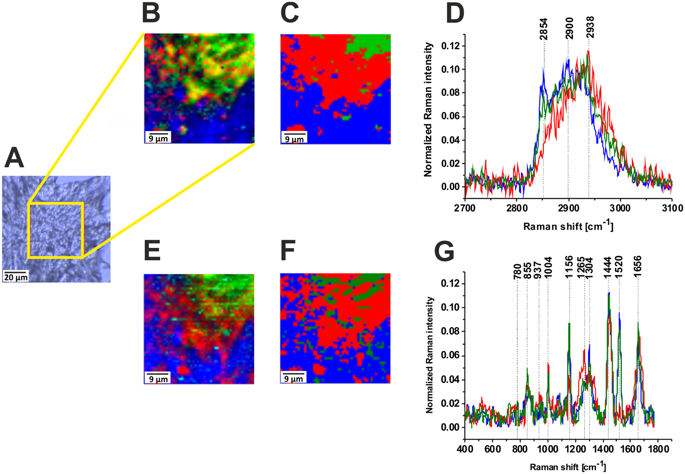
We have shown that Raman microspectroscopy is a powerful method for visualization of glycocalyx offering cellular interrogation without staining, unprecedented spatial and spectral resolution, and biochemical information. We showed for the first time that Raman imaging can be used to distinguish successfully between glycosylated and nonglycosylated proteins in normal and cancer tissue. Thousands of protein, lipid and glycan species exist in cells and tissues and their metabolism is monitored via numerous pathways, networks and methods. The metabolism can change in response to cellular environment alterations, such as development of a disease. Measuring such alterations and understanding the pathways involved are crucial to fully understand cellular metabolism in cancer development. In this paper Raman markers of glycogen, glycosaminoglycan, chondroitin sulfate, heparan sulfate proteoglycan were identified based on their vibrational signatures. High spatial resolution of Raman imaging combined with chemometrics allows separation of individual species from many chemical components present in each cell. We have found that metabolism of proteins, lipids and glycans is markedly deregulated in breast (adenocarcinoma) and brain (medulloblastoma) tumors. We have identified two glycoforms in the normal breast tissue and the malignant brain tissue in contrast to the breast cancer tissue where only one glycoform has been identified.
中文翻译:

通过拉曼成像监测脑癌和乳腺癌的糖基化代谢。
我们已经证明,拉曼显微光谱是一种强大的糖萼可视化方法,可提供无需染色的细胞询问、前所未有的空间和光谱分辨率以及生化信息。我们首次证明拉曼成像可用于成功区分正常组织和癌症组织中的糖基化和非糖基化蛋白质。细胞和组织中存在数千种蛋白质、脂质和聚糖,它们的代谢通过多种途径、网络和方法进行监测。新陈代谢可以响应细胞环境的变化而改变,例如疾病的发展。测量此类改变并了解所涉及的途径对于充分了解癌症发展中的细胞代谢至关重要。在本文中,根据糖原、糖胺聚糖、硫酸软骨素、硫酸乙酰肝素蛋白聚糖的拉曼标记物的振动特征进行了鉴定。拉曼成像的高空间分辨率与化学计量学相结合,可以将单个物种与每个细胞中存在的许多化学成分分离。我们发现,在乳腺癌(腺癌)和脑肿瘤(髓母细胞瘤)中,蛋白质、脂质和聚糖的代谢明显失调。我们在正常乳腺组织和恶性脑组织中鉴定出两种糖型,而在乳腺癌组织中仅鉴定出一种糖型。
更新日期:2019-01-17
中文翻译:

通过拉曼成像监测脑癌和乳腺癌的糖基化代谢。
我们已经证明,拉曼显微光谱是一种强大的糖萼可视化方法,可提供无需染色的细胞询问、前所未有的空间和光谱分辨率以及生化信息。我们首次证明拉曼成像可用于成功区分正常组织和癌症组织中的糖基化和非糖基化蛋白质。细胞和组织中存在数千种蛋白质、脂质和聚糖,它们的代谢通过多种途径、网络和方法进行监测。新陈代谢可以响应细胞环境的变化而改变,例如疾病的发展。测量此类改变并了解所涉及的途径对于充分了解癌症发展中的细胞代谢至关重要。在本文中,根据糖原、糖胺聚糖、硫酸软骨素、硫酸乙酰肝素蛋白聚糖的拉曼标记物的振动特征进行了鉴定。拉曼成像的高空间分辨率与化学计量学相结合,可以将单个物种与每个细胞中存在的许多化学成分分离。我们发现,在乳腺癌(腺癌)和脑肿瘤(髓母细胞瘤)中,蛋白质、脂质和聚糖的代谢明显失调。我们在正常乳腺组织和恶性脑组织中鉴定出两种糖型,而在乳腺癌组织中仅鉴定出一种糖型。


 京公网安备 11010802027423号