当前位置:
X-MOL 学术
›
Adv. Funct. Mater.
›
论文详情
Our official English website, www.x-mol.net, welcomes your
feedback! (Note: you will need to create a separate account there.)
[4]Helicene–Squalene Fluorescent Nanoassemblies for Specific Targeting of Mitochondria in Live‐Cell Imaging
Advanced Functional Materials ( IF 18.5 ) Pub Date : 2017-07-10 , DOI: 10.1002/adfm.201701839 Andrej Babič 1 , Simon Pascal 2 , Romain Duwald 2 , Dimitri Moreau 3 , Jérôme Lacour 2 , Eric Allémann 1
Advanced Functional Materials ( IF 18.5 ) Pub Date : 2017-07-10 , DOI: 10.1002/adfm.201701839 Andrej Babič 1 , Simon Pascal 2 , Romain Duwald 2 , Dimitri Moreau 3 , Jérôme Lacour 2 , Eric Allémann 1
Affiliation

|
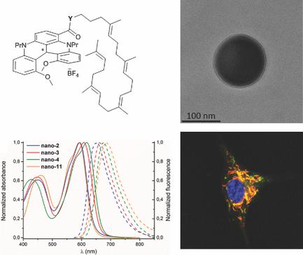
Ester, amide, and directly linked composites of squalene and cationic diaza [4]helicenes 1 are readily prepared. These lipid‐dye constructs 2, 3, and 4 give in aqueous media monodispersed spherical nanoassemblies around 100–130 nm in diameter with excellent stability for several months. Racemic and enantiopure nanoassemblies of compound 2 are fully characterized, including by transmission electron microscope and cryogenic transmission electron microscope imaging that did not reveal higher order supramolecular structures. Investigations of their (chir)optical properties show red absorption maxima ≈600 nm and red fluorescence spanning up to the near‐infrared region, with average Stokes shifts of 1350–1550 cm−1. Live‐cell imaging by confocal microscopy reveals rapid internalization on the minute time scale and organelle‐specific accumulation. Colocalization with MitoTracker in several cancer cell lines demonstrates a specific staining of mitochondria by the [4]helicene–squalene nanoassemblies. To our knowledge, it is the first report of a subcellular targeting by squalene‐based nanoassemblies.
中文翻译:

[4]用于活细胞成像中线粒体特异性靶向的螺旋烯-角鲨烯荧光纳米组装体
角鲨烯和阳离子二氮杂[4]螺旋烯1的酯,酰胺和直接连接的复合物易于制备。这些脂质染料构建体2,3和4给予在水介质中的单分散球形nanoassemblies周围100-130纳米的直径具有优良的稳定性数月。化合物2的外消旋和对映纯纳米组装件得到了充分表征,包括通过透射电子显微镜和低温透射电子显微镜成像,而这些成像都没有揭示高阶超分子结构。对它们的(手性)光学特性的研究表明,最大吸收峰≈600nm,红色荧光跨越近红外区域,平均斯托克斯位移为1350–1550 cm-1。通过共聚焦显微镜对活细胞成像显示出在微小的时间范围内的快速内在化和特定于细胞器的积累。与MitoTracker在几种癌细胞系中的共定位显示了[4]螺旋烯-角鲨烯纳米组装体对线粒体的特异性染色。据我们所知,这是基于角鲨烯的纳米组件靶向亚细胞的首次报道。
更新日期:2017-07-10
中文翻译:

[4]用于活细胞成像中线粒体特异性靶向的螺旋烯-角鲨烯荧光纳米组装体
角鲨烯和阳离子二氮杂[4]螺旋烯1的酯,酰胺和直接连接的复合物易于制备。这些脂质染料构建体2,3和4给予在水介质中的单分散球形nanoassemblies周围100-130纳米的直径具有优良的稳定性数月。化合物2的外消旋和对映纯纳米组装件得到了充分表征,包括通过透射电子显微镜和低温透射电子显微镜成像,而这些成像都没有揭示高阶超分子结构。对它们的(手性)光学特性的研究表明,最大吸收峰≈600nm,红色荧光跨越近红外区域,平均斯托克斯位移为1350–1550 cm-1。通过共聚焦显微镜对活细胞成像显示出在微小的时间范围内的快速内在化和特定于细胞器的积累。与MitoTracker在几种癌细胞系中的共定位显示了[4]螺旋烯-角鲨烯纳米组装体对线粒体的特异性染色。据我们所知,这是基于角鲨烯的纳米组件靶向亚细胞的首次报道。


 京公网安备 11010802027423号