当前位置:
X-MOL 学术
›
Hum. Brain Mapp.
›
论文详情
Our official English website, www.x-mol.net, welcomes your
feedback! (Note: you will need to create a separate account there.)
Automatic detection of neuromelanin and iron in the midbrain nuclei using a magnetic resonance imaging-based brain template
Human Brain Mapping ( IF 3.5 ) Pub Date : 2022-01-24 , DOI: 10.1002/hbm.25770 Zhijia Jin 1 , Ying Wang 2, 3 , Mojtaba Jokar 2 , Yan Li 1 , Zenghui Cheng 1 , Yu Liu 1 , Rongbiao Tang 1 , Xiaofeng Shi 1 , Youmin Zhang 1 , Jihua Min 1 , Fangtao Liu 1 , Naying He 1 , Fuhua Yan 1 , Ewart Mark Haacke 1, 2, 3, 4, 5
Human Brain Mapping ( IF 3.5 ) Pub Date : 2022-01-24 , DOI: 10.1002/hbm.25770 Zhijia Jin 1 , Ying Wang 2, 3 , Mojtaba Jokar 2 , Yan Li 1 , Zenghui Cheng 1 , Yu Liu 1 , Rongbiao Tang 1 , Xiaofeng Shi 1 , Youmin Zhang 1 , Jihua Min 1 , Fangtao Liu 1 , Naying He 1 , Fuhua Yan 1 , Ewart Mark Haacke 1, 2, 3, 4, 5
Affiliation

|
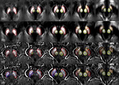
Parkinson disease (PD) is a chronic progressive neurodegenerative disorder characterized pathologically by early loss of neuromelanin (NM) in the substantia nigra pars compacta (SNpc) and increased iron deposition in the substantia nigra (SN). Degeneration of the SN presents as a 50 to 70% loss of pigmented neurons in the ventral lateral tier of the SNpc at the onset of symptoms. Also, using magnetic resonance imaging (MRI), iron deposition and volume changes of the red nucleus (RN), and subthalamic nucleus (STN) have been reported to be associated with disease status and rate of progression. Further, the STN serves as an important target for deep brain stimulation treatment in advanced PD patients. Therefore, an accurate in-vivo delineation of the SN, its subregions and other midbrain structures such as the RN and STN could be useful to better study iron and NM changes in PD. Our goal was to use an MRI template to create an automatic midbrain deep gray matter nuclei segmentation approach based on iron and NM contrast derived from a single, multiecho magnetization transfer contrast gradient echo (MTC-GRE) imaging sequence. The short echo TE = 7.5 ms data from a 3D MTC-GRE sequence was used to find the NM-rich region, while the second echo TE = 15 ms was used to calculate the quantitative susceptibility map for 87 healthy subjects (mean age ± SD: 63.4 ± 6.2 years old, range: 45–81 years). From these data, we created both NM and iron templates and calculated the boundaries of each midbrain nucleus in template space, mapped these boundaries back to the original space and then fine-tuned the boundaries in the original space using a dynamic programming algorithm to match the details of each individual's NM and iron features. A dual mapping approach was used to improve the performance of the morphological mapping of the midbrain of any given individual to the template space. A threshold approach was used in the NM-rich region and susceptibility maps to optimize the DICE similarity coefficients and the volume ratios. The results for the NM of the SN as well as the iron containing SN, STN, and RN all indicate a strong agreement with manually drawn structures. The DICE similarity coefficients and volume ratios for these structures were 0.85, 0.87, 0.75, and 0.92 and 0.93, 0.95, 0.89, 1.05, respectively, before applying any threshold on the data. Using this fully automatic template-based deep gray matter mapping approach, it is possible to accurately measure the tissue properties such as volumes, iron content, and NM content of the midbrain nuclei.
中文翻译:

使用基于磁共振成像的脑模板自动检测中脑核中的神经黑色素和铁
帕金森病 (PD) 是一种慢性进行性神经退行性疾病,其病理特征是黑质致密部 (SNpc) 中神经黑色素 (NM) 的早期丧失和黑质 (SN) 中铁沉积增加。SN 退化表现为症状发作时 SNpc 腹外侧层的色素神经元损失 50% 至 70%。此外,据报道,使用磁共振成像 (MRI),红核 (RN) 和丘脑底核 (STN) 的铁沉积和体积变化与疾病状态和进展速度有关。此外,STN 是晚期 PD 患者深部脑刺激治疗的重要靶点。因此,SN 的准确体内描绘,其子区域和其他中脑结构(如 RN 和 STN)可能有助于更好地研究 PD 中的铁和 NM 变化。我们的目标是使用 MRI 模板创建基于铁和 NM 对比度的自动中脑深部灰质核分割方法,该方法源自单个多回波磁化转移对比梯度回波 (MTC-GRE) 成像序列。来自 3D MTC-GRE 序列的短回波 TE = 7.5 ms 数据用于寻找 NM 丰富区域,而第二个回波 TE = 15 ms 用于计算 87 名健康受试者(平均年龄 ± 多回波磁化转移对比梯度回波 (MTC-GRE) 成像序列。来自 3D MTC-GRE 序列的短回波 TE = 7.5 ms 数据用于寻找 NM 丰富区域,而第二个回波 TE = 15 ms 用于计算 87 名健康受试者(平均年龄 ± 多回波磁化转移对比梯度回波 (MTC-GRE) 成像序列。来自 3D MTC-GRE 序列的短回波 TE = 7.5 ms 数据用于寻找 NM 丰富区域,而第二个回波 TE = 15 ms 用于计算 87 名健康受试者(平均年龄 ± 标清:63.4 ± 6.2 岁,范围:45-81 岁)。根据这些数据,我们创建了 NM 和铁模板,并计算了模板空间中每个中脑核的边界,将这些边界映射回原始空间,然后使用动态规划算法微调原始空间中的边界以匹配每个人的 NM 和铁特征的详细信息。使用双重映射方法来提高任何给定个体的中脑到模板空间的形态映射性能。在富含 NM 的区域和敏感性图中使用阈值方法来优化 DICE 相似性系数和体积比。SN 的 NM 以及含铁的 SN、STN 和 RN 的结果都表明与手工绘制的结构有很强的一致性。在对数据应用任何阈值之前,这些结构的 DICE 相似性系数和体积比分别为 0.85、0.87、0.75 和 0.92 和 0.93、0.95、0.89、1.05。使用这种基于模板的全自动深灰质映射方法,可以准确测量组织特性,例如中脑核的体积、铁含量和 NM 含量。
更新日期:2022-01-24
中文翻译:

使用基于磁共振成像的脑模板自动检测中脑核中的神经黑色素和铁
帕金森病 (PD) 是一种慢性进行性神经退行性疾病,其病理特征是黑质致密部 (SNpc) 中神经黑色素 (NM) 的早期丧失和黑质 (SN) 中铁沉积增加。SN 退化表现为症状发作时 SNpc 腹外侧层的色素神经元损失 50% 至 70%。此外,据报道,使用磁共振成像 (MRI),红核 (RN) 和丘脑底核 (STN) 的铁沉积和体积变化与疾病状态和进展速度有关。此外,STN 是晚期 PD 患者深部脑刺激治疗的重要靶点。因此,SN 的准确体内描绘,其子区域和其他中脑结构(如 RN 和 STN)可能有助于更好地研究 PD 中的铁和 NM 变化。我们的目标是使用 MRI 模板创建基于铁和 NM 对比度的自动中脑深部灰质核分割方法,该方法源自单个多回波磁化转移对比梯度回波 (MTC-GRE) 成像序列。来自 3D MTC-GRE 序列的短回波 TE = 7.5 ms 数据用于寻找 NM 丰富区域,而第二个回波 TE = 15 ms 用于计算 87 名健康受试者(平均年龄 ± 多回波磁化转移对比梯度回波 (MTC-GRE) 成像序列。来自 3D MTC-GRE 序列的短回波 TE = 7.5 ms 数据用于寻找 NM 丰富区域,而第二个回波 TE = 15 ms 用于计算 87 名健康受试者(平均年龄 ± 多回波磁化转移对比梯度回波 (MTC-GRE) 成像序列。来自 3D MTC-GRE 序列的短回波 TE = 7.5 ms 数据用于寻找 NM 丰富区域,而第二个回波 TE = 15 ms 用于计算 87 名健康受试者(平均年龄 ± 标清:63.4 ± 6.2 岁,范围:45-81 岁)。根据这些数据,我们创建了 NM 和铁模板,并计算了模板空间中每个中脑核的边界,将这些边界映射回原始空间,然后使用动态规划算法微调原始空间中的边界以匹配每个人的 NM 和铁特征的详细信息。使用双重映射方法来提高任何给定个体的中脑到模板空间的形态映射性能。在富含 NM 的区域和敏感性图中使用阈值方法来优化 DICE 相似性系数和体积比。SN 的 NM 以及含铁的 SN、STN 和 RN 的结果都表明与手工绘制的结构有很强的一致性。在对数据应用任何阈值之前,这些结构的 DICE 相似性系数和体积比分别为 0.85、0.87、0.75 和 0.92 和 0.93、0.95、0.89、1.05。使用这种基于模板的全自动深灰质映射方法,可以准确测量组织特性,例如中脑核的体积、铁含量和 NM 含量。


 京公网安备 11010802027423号