Our official English website, www.x-mol.net, welcomes your
feedback! (Note: you will need to create a separate account there.)
Label-Free Dynamic Imaging of Chromatin in Live Cell Nuclei by High-Speed Scattering-Based Interference Microscopy
ACS Nano ( IF 15.8 ) Pub Date : 2021-12-30 , DOI: 10.1021/acsnano.1c09748 Yi-Teng Hsiao, Chia-Ni Tsai, Te-Hsin Chen, Chia-Lung Hsieh
ACS Nano ( IF 15.8 ) Pub Date : 2021-12-30 , DOI: 10.1021/acsnano.1c09748 Yi-Teng Hsiao, Chia-Ni Tsai, Te-Hsin Chen, Chia-Lung Hsieh

|
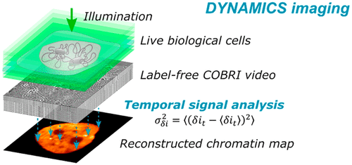
Chromatin is a DNA–protein complex that is densely packed in the cell nucleus. The nanoscale chromatin compaction plays critical roles in the modulation of cell nuclear processes. However, little is known about the spatiotemporal dynamics of chromatin compaction states because it remains difficult to quantitatively measure the chromatin compaction level in live cells. Here, we demonstrate a strategy, referenced as DYNAMICS imaging, for mapping chromatin organization in live cell nuclei by analyzing the dynamic scattering signal of molecular fluctuations. Highly sensitive optical interference microscopy, coherent brightfield (COBRI) microscopy, is implemented to detect the linear scattering of unlabeled chromatin at a high speed. A theoretical model is established to determine the local chromatin density from the statistical fluctuation of the measured scattering signal. DYNAMICS imaging allows us to reconstruct a speckle-free nucleus map that is highly correlated to the fluorescence chromatin image. Moreover, together with calibration based on nanoparticle colloids, we show that the DYNAMICS signal is sensitive to the chromatin compaction level at the nanoscale. We confirm the effectiveness of DYNAMICS imaging in detecting the condensation and decondensation of chromatin induced by chemical drug treatments. Importantly, the stable scattering signal supports a continuous observation of the chromatin condensation and decondensation processes for more than 1 h. Using this technique, we detect transient and nanoscopic chromatin condensation events occurring on a time scale of a few seconds. Label-free DYNAMICS imaging offers the opportunity to investigate chromatin conformational dynamics and to explore their significance in various gene activities.
中文翻译:

基于高速散射的干涉显微镜对活细胞核中染色质的无标记动态成像
染色质是一种紧密堆积在细胞核中的 DNA-蛋白质复合物。纳米级染色质压实在细胞核过程的调节中起着关键作用。然而,对染色质压实状态的时空动态知之甚少,因为仍然难以定量测量活细胞中的染色质压实水平。在这里,我们展示了一种称为 DYNAMICS 成像的策略,用于通过分析分子波动的动态散射信号来绘制活细胞核中的染色质组织。高灵敏度光学干涉显微镜,相干明场 (COBRI) 显微镜,用于高速检测未标记染色质的线性散射。建立了一个理论模型,从测量的散射信号的统计波动中确定局部染色质密度。DYNAMICS 成像使我们能够重建与荧光染色质图像高度相关的无斑点核图。此外,结合基于纳米颗粒胶体的校准,我们表明 DYNAMICS 信号对纳米尺度的染色质压实水平敏感。我们证实了 DYNAMICS 成像在检测化学药物处理引起的染色质凝聚和去凝聚方面的有效性。重要的是,稳定的散射信号支持对染色质凝聚和去凝聚过程的连续观察超过 1 小时。使用这种技术,我们检测到在几秒钟的时间尺度上发生的瞬态和纳米级染色质凝聚事件。无标记动态成像提供了研究染色质构象动力学并探索其在各种基因活动中的意义的机会。
更新日期:2021-12-30
中文翻译:

基于高速散射的干涉显微镜对活细胞核中染色质的无标记动态成像
染色质是一种紧密堆积在细胞核中的 DNA-蛋白质复合物。纳米级染色质压实在细胞核过程的调节中起着关键作用。然而,对染色质压实状态的时空动态知之甚少,因为仍然难以定量测量活细胞中的染色质压实水平。在这里,我们展示了一种称为 DYNAMICS 成像的策略,用于通过分析分子波动的动态散射信号来绘制活细胞核中的染色质组织。高灵敏度光学干涉显微镜,相干明场 (COBRI) 显微镜,用于高速检测未标记染色质的线性散射。建立了一个理论模型,从测量的散射信号的统计波动中确定局部染色质密度。DYNAMICS 成像使我们能够重建与荧光染色质图像高度相关的无斑点核图。此外,结合基于纳米颗粒胶体的校准,我们表明 DYNAMICS 信号对纳米尺度的染色质压实水平敏感。我们证实了 DYNAMICS 成像在检测化学药物处理引起的染色质凝聚和去凝聚方面的有效性。重要的是,稳定的散射信号支持对染色质凝聚和去凝聚过程的连续观察超过 1 小时。使用这种技术,我们检测到在几秒钟的时间尺度上发生的瞬态和纳米级染色质凝聚事件。无标记动态成像提供了研究染色质构象动力学并探索其在各种基因活动中的意义的机会。


 京公网安备 11010802027423号