Our official English website, www.x-mol.net, welcomes your
feedback! (Note: you will need to create a separate account there.)
Single-Cell Mass Spectrometry Imaging of Multiple Drugs and Nanomaterials at Organelle Level
ACS Nano ( IF 15.8 ) Pub Date : 2021-07-27 , DOI: 10.1021/acsnano.1c02922 Yifan Meng 1 , Chaohong Gao 1 , Qiao Lu 1 , Siyuan Ma 1 , Wei Hang 1
ACS Nano ( IF 15.8 ) Pub Date : 2021-07-27 , DOI: 10.1021/acsnano.1c02922 Yifan Meng 1 , Chaohong Gao 1 , Qiao Lu 1 , Siyuan Ma 1 , Wei Hang 1
Affiliation

|
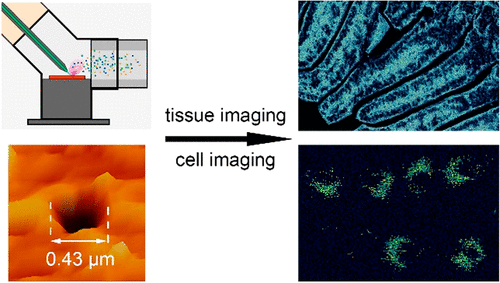
Mass spectrometry imaging (MSI) techniques make possible the spatial chemical identification of analytes, especially for biological samples. As a universal energy source, laser is one of the most commonly used sampling methods in MSI techniques. However, due to the limitation of laser spot size, subcellular spatial resolution imaging, which is significant for life science researches, always remains a challenge for laser-based MSI. In this research, we designed a laser ablation (LA) system with a microlensed fiber and a “three-way” structure ablation chamber, and achieved nanoscale inductively coupled plasma (ICP) MSI with an adjustable spatial resolution down to 400 nm, which surpasses most existing technologies. With this device, the distribution of various photodynamic therapy drugs in the intestine of mouse can be clearly observed. The comparison imaging results showed that the drug distribution in tissue slice could be identified at the subcellular level with the high-resolution mode. More valuably, gold nanorods (GNRs) and carboplatin in a single cell are able to be visualized at organelle level due to the nanoscale resolution, which is able to reveal the mechanism of cell apoptosis. This reliable and economical MSI technique is expected to be used in understanding the precise chemical composition and transportation in small tissues, microorganisms, and single cells.
中文翻译:

多种药物和纳米材料在细胞器水平的单细胞质谱成像
质谱成像 (MSI) 技术使分析物的空间化学鉴定成为可能,尤其是对于生物样品。作为一种通用能源,激光是MSI技术中最常用的采样方法之一。然而,由于激光光斑尺寸的限制,对生命科学研究具有重要意义的亚细胞空间分辨率成像一直是激光微星成像面临的挑战。在这项研究中,我们设计了一种具有微透镜光纤和“三通”结构消融室的激光消融(LA)系统,实现了空间分辨率可调至400 nm的纳米级电感耦合等离子体(ICP)MSI,超过了大多数现有技术。通过该装置,可以清楚地观察到各种光动力治疗药物在小鼠肠道内的分布情况。对比成像结果表明,在高分辨率模式下,可以在亚细胞水平上识别组织切片中的药物分布。更有价值的是,由于纳米级分辨率,单个细胞中的金纳米棒 (GNR) 和卡铂能够在细胞器水平上被可视化,这能够揭示细胞凋亡的机制。这种可靠且经济的 MSI 技术有望用于了解小组织、微生物和单细胞中精确的化学成分和运输。能够揭示细胞凋亡的机制。这种可靠且经济的 MSI 技术有望用于了解小组织、微生物和单细胞中精确的化学成分和运输。能够揭示细胞凋亡的机制。这种可靠且经济的 MSI 技术有望用于了解小组织、微生物和单细胞中精确的化学成分和运输。
更新日期:2021-08-24
中文翻译:

多种药物和纳米材料在细胞器水平的单细胞质谱成像
质谱成像 (MSI) 技术使分析物的空间化学鉴定成为可能,尤其是对于生物样品。作为一种通用能源,激光是MSI技术中最常用的采样方法之一。然而,由于激光光斑尺寸的限制,对生命科学研究具有重要意义的亚细胞空间分辨率成像一直是激光微星成像面临的挑战。在这项研究中,我们设计了一种具有微透镜光纤和“三通”结构消融室的激光消融(LA)系统,实现了空间分辨率可调至400 nm的纳米级电感耦合等离子体(ICP)MSI,超过了大多数现有技术。通过该装置,可以清楚地观察到各种光动力治疗药物在小鼠肠道内的分布情况。对比成像结果表明,在高分辨率模式下,可以在亚细胞水平上识别组织切片中的药物分布。更有价值的是,由于纳米级分辨率,单个细胞中的金纳米棒 (GNR) 和卡铂能够在细胞器水平上被可视化,这能够揭示细胞凋亡的机制。这种可靠且经济的 MSI 技术有望用于了解小组织、微生物和单细胞中精确的化学成分和运输。能够揭示细胞凋亡的机制。这种可靠且经济的 MSI 技术有望用于了解小组织、微生物和单细胞中精确的化学成分和运输。能够揭示细胞凋亡的机制。这种可靠且经济的 MSI 技术有望用于了解小组织、微生物和单细胞中精确的化学成分和运输。


 京公网安备 11010802027423号