Journal of Ethnopharmacology ( IF 4.8 ) Pub Date : 2021-05-06 , DOI: 10.1016/j.jep.2021.114190 Rui Li 1 , Xiaopeng Ai 1 , Ya Hou 2 , Xianrong Lai 3 , Xianli Meng 4 , Xiaobo Wang 2

|
Ethnopharmacological relevance
Berberis dictyophylla F., a famous Tibetan medicine, has been used to prevent and treat diabetic retinopathy (DR) for thousands of years in clinic. However, its underlying mechanisms remain unclear.
Aim of the study
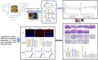
The present study was designed to probe the synergistic protection and involved mechanisms of berberine, magnoflorine and berbamine from Berberis dictyophylla F. on the spontaneous retinal damage of db/db mice.
Materials and methods
The 14-week spontaneous model of DR in db/db mice were randomly divided into eight groups: model group, calcium dobesilate (CaDob, 0.23 g/kg) group and groups 1–6 (different proportional three active ingredients from Berberis dictyophylla F.). All mice were intragastrically administrated for a continuous 12 weeks. Body weight and fasting blood glucose (FBG) were recorded and measured. Hematoxylin-eosin and periodic acid–Schiff (PAS) stainings were employed to evaluate the pathological changes and abnormal angiogenesis of the retina. ELISA was performed to assess the levels of IL-6, HIF-1α and VEGF in the serum. Immunofluorescent staining was applied to detect the protein levels of CD31, VEGF, p-p38, p-JNK, p-ERK and NF-κB in retina. In addition, mRNA expression levels of VEGF, Bax and Bcl-2 in the retina were monitored by qRT-PCR analysis.
Results
Treatment with different proportional three active ingredients exerted no significant effect on the weight, but decreased the FBG, increased the number of retinal ganglionic cells and restored internal limiting membrane. The results of PAS staining demonstrated that the drug treatment decreased the ratio of endothelial cells to pericytes while thinned the basal membrane of retinal vessels. Moreover, these different proportional active ingredients can markedly downregulate the protein levels of retinal CD31 and VEGF, and serum HIF-1α and VEGF. The gene expression of retinal VEGF was also suppressed. The levels of retinal p-p38, p-JNK and p-ERK proteins were decreased by drug treatment. Finally, drug treatment reversed the proinflammatory factors of retinal NF-κB and serum IL-6, and proapoptotic Bax gene expression, while increased antiapoptotic Bcl-2 gene expression.
Conclusions
These results indicated that DR in db/db mice can be ameliorated by treatment with different proportional three active ingredients from Berberis dictyophylla F. The potential vascular protection mechanisms may be involved in inhibiting the phosphorylation of the MAPK signaling pathway, thus decreasing inflammatory and apoptotic events.
中文翻译:

藏药小檗F三种活性成分不同比例治疗db/db小鼠糖尿病视网膜病变
民族药理学相关性
小檗是著名的藏药,在临床上用于预防和治疗糖尿病视网膜病变(DR)已有数千年的历史。然而,其潜在机制仍不清楚。
研究目的
本研究旨在探讨小檗碱、木兰花碱和小檗胺对db/db小鼠自发性视网膜损伤的协同保护作用及其机制。
材料和方法
db/db小鼠14周自发DR模型随机分为8组:模型组、羟苯磺酸钙(CaDob,0.23 g/kg)组和1-6组(不同比例的小檗中的三种活性成分)。 )。所有小鼠均连续灌胃给药12周。记录并测量体重和空腹血糖(FBG)。采用苏木精-伊红和高碘酸-希夫(PAS)染色来评估视网膜的病理变化和异常血管生成。采用ELISA法评估血清中IL-6、HIF-1α和VEGF的水平。免疫荧光染色检测视网膜CD31、VEGF、p-p38、p-JNK、p-ERK、NF-κB蛋白水平。此外,通过qRT-PCR分析监测视网膜中VEGF、Bax和Bcl-2的mRNA表达水平。
结果
不同比例的三种活性成分治疗对体重无明显影响,但FBG下降,视网膜神经节细胞数量增加,内界膜恢复。 PAS染色结果表明,药物治疗降低了内皮细胞与周细胞的比例,同时使视网膜血管基底膜变薄。而且,这些不同比例的活性成分能够显着下调视网膜CD31和VEGF以及血清HIF-1α和VEGF的蛋白水平。视网膜VEGF的基因表达也受到抑制。药物治疗降低了视网膜 p-p38、p-JNK 和 p-ERK 蛋白的水平。最后,药物治疗逆转了视网膜NF-κB和血清IL-6的促炎因子以及促凋亡Bax基因的表达,同时增加了抗凋亡Bcl-2基因的表达。
结论
这些结果表明,用不同比例的网叶小檗三种活性成分治疗可以改善db/db小鼠的DR。潜在的血管保护机制可能涉及抑制MAPK信号通路的磷酸化,从而减少炎症和细胞凋亡事件。


 京公网安备 11010802027423号