当前位置:
X-MOL 学术
›
J. Mater. Chem. B
›
论文详情
Our official English website, www.x-mol.net, welcomes your
feedback! (Note: you will need to create a separate account there.)
NIR-I-to-NIR-II fluorescent nanomaterials for biomedical imaging and cancer therapy
Journal of Materials Chemistry B ( IF 6.1 ) Pub Date : 2017-11-23 00:00:00 , DOI: 10.1039/c7tb02573d Jingya Zhao 1, 2, 3, 4, 5 , Dian Zhong 1, 2, 3, 4, 5 , Shaobing Zhou 1, 2, 3, 4, 5
Journal of Materials Chemistry B ( IF 6.1 ) Pub Date : 2017-11-23 00:00:00 , DOI: 10.1039/c7tb02573d Jingya Zhao 1, 2, 3, 4, 5 , Dian Zhong 1, 2, 3, 4, 5 , Shaobing Zhou 1, 2, 3, 4, 5
Affiliation

|
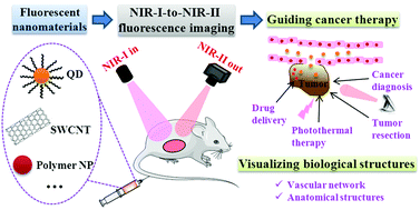
Near-infrared (NIR) fluorescence imaging, which affords high imaging resolution owing to deep tissue penetration of NIR photons, is an attractive imaging modality for both biomedical research and clinical applications. To further improve the image contrast at increased tissue depth, recently much attention has been focused on the development of NIR-I-to-NIR-II fluorescence imaging, which can remarkably reduce the interference from photon absorption, scattering and tissue autofluorescence with excitation in the 700–950 nm NIR-I window and emission in the 1000–1700 nm NIR-II window. In this review, we highlight recently developed NIR-I-to-NIR-II fluorescent nanomaterials, including silver chalcogenide quantum dots, single-walled carbon nanotubes and polymer nanoparticles. We discuss the advantages of these nanomaterials as fluorescent tags in deep tissue imaging by comparing them with conventional fluorophores, and then survey the implementation of NIR fluorescence imaging with these nanomaterials, including instrumentation, data analysis and surface biofunctionalization of the nanomaterials. Finally, we discuss recent applications of NIR-I-to-NIR-II fluorescent nanomaterials in the biomedical imaging field, with an emphasis on how to use them to achieve simultaneous cancer diagnosis and therapy.
中文翻译:

用于生物医学成像和癌症治疗的NIR-I至NIR-II荧光纳米材料
近红外(NIR)荧光成像由于对NIR光子的深层组织穿透而提供了很高的成像分辨率,对于生物医学研究和临床应用而言都是有吸引力的成像方式。为了进一步提高在组织深度增加时的图像对比度,近来已将许多注意力集中在NIR-I至NIR-II荧光成像的开发上,该成像可以显着降低光子吸收,散射和组织自发荧光在激发下的干扰。 700–950 nm NIR-I窗口和1000–1700 nm NIR-II窗口中的发射。在这篇评论中,我们重点介绍了最近开发的NIR-I至NIR-II荧光纳米材料,包括硫属元素化银量子点,单壁碳纳米管和聚合物纳米颗粒。通过与常规荧光团进行比较,我们讨论了这些纳米材料作为荧光标签在深部组织成像中的优势,然后调查了这些纳米材料在近红外荧光成像中的应用,包括纳米材料的仪器,数据分析和表面生物功能化。最后,我们讨论了NIR-I至NIR-II荧光纳米材料在生物医学成像领域的最新应用,重点是如何使用它们来实现同时的癌症诊断和治疗。
更新日期:2017-11-23
中文翻译:

用于生物医学成像和癌症治疗的NIR-I至NIR-II荧光纳米材料
近红外(NIR)荧光成像由于对NIR光子的深层组织穿透而提供了很高的成像分辨率,对于生物医学研究和临床应用而言都是有吸引力的成像方式。为了进一步提高在组织深度增加时的图像对比度,近来已将许多注意力集中在NIR-I至NIR-II荧光成像的开发上,该成像可以显着降低光子吸收,散射和组织自发荧光在激发下的干扰。 700–950 nm NIR-I窗口和1000–1700 nm NIR-II窗口中的发射。在这篇评论中,我们重点介绍了最近开发的NIR-I至NIR-II荧光纳米材料,包括硫属元素化银量子点,单壁碳纳米管和聚合物纳米颗粒。通过与常规荧光团进行比较,我们讨论了这些纳米材料作为荧光标签在深部组织成像中的优势,然后调查了这些纳米材料在近红外荧光成像中的应用,包括纳米材料的仪器,数据分析和表面生物功能化。最后,我们讨论了NIR-I至NIR-II荧光纳米材料在生物医学成像领域的最新应用,重点是如何使用它们来实现同时的癌症诊断和治疗。


 京公网安备 11010802027423号